, Anna Maria Belli2 , Joo-Young Chun3, Raymond Chung3, Raj Das3, Andrew England4, Karen Flood5, Marie-France Giroux6, Richard G. McWilliams7, Robert Morgan3, Nik Papadakos3, Jai V. Patel8, Raf Patel8 , Uday Patel9 , Lakshmi Ratnam10 , Reddi Prasad Yadavali11 and John Rose12
(1)
Department of Interventional Radiology, University Hospitals Southampton, Southampton, Hampshire, UK
(2)
Department of Radiology, St. George’s Hospital and Medical School, Blackshaw Road, London, SW17 0RE, UK
(3)
Department of Radiology, St. George’s Hospital, London, UK
(4)
Department of Radiography, University of Salford, Manchester, UK
(5)
Department of Vascular Radiology, Leeds General Infirmary, Leeds, UK
(6)
Department of Radiology, CHUM-Centre Hospitalier de l’Université de Montréal, Montreal, QC, Canada
(7)
Department of Radiology, Royal Liverpool University Hospital, Liverpool, UK
(8)
Department of Radiology, The Leeds Teaching Hospitals NHS Trust, Leeds, West Yorkshire, UK
(9)
Department of Diagnostic Radiology, St. George’s Hospital and Medical School, Blackshaw Road, SW17 0QT London, UK
(10)
Department of Radiology, St. George’s Hospital, Blackshaw Road, SW17 0QT London, UK
(11)
Department of Radiology, Aberdeen Royal Infirmary, Aberdeen, UK
(12)
Department of Interventional Radiology, Freeman Hospital, Newcastle Upon Tyne Hospitals NHS Trust, Newcastle upon Tyne, UK
Abstract
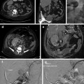
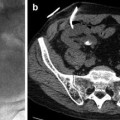
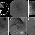
This case reviews the different approaches to dealing with acute thrombosis following angioplasty. An example of a thrombolysis regimen is given in addition to technique for thromboaspiration.
Keywords
ComplicationsAngioplastyThrombosisThrombolysisCase History
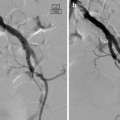
A morbidly obese 58-year-old male presented with rest pain in his left foot. His other relevant medical history was of insulin-dependent diabetes, hypertension, and hyperlipidemia. The patient was too large to be imaged in the departmental MRI scanner. A lower limb Doppler ultrasound examination demonstrated significant mid-left SFA stenosis, and angioplasty of this lesion was planned.
