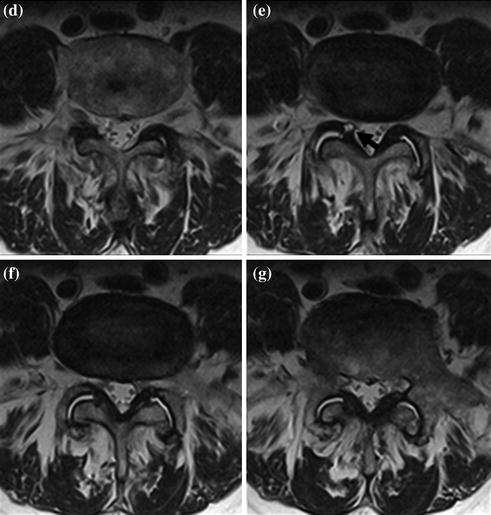
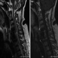
Fig. 1
a–g. FSE T2 sagittal (a–c) and axial (d–g) sections. L4-L5 small cystic lesion in right foraminal recess (arrow), supplied by inter-apophyseal fluid. Bilateral interapophyseal degenerative phenomenon, with articular hypertrophy and subsequent narrowing of vertebral canal (e–g)
Pre-operative Imaging
 Herniated Lumbar Disk Diskectomy
 Dorsal Herniated Disk Diskectomy and Stabilization
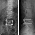 Lumbar Stenosis and Degenerative Instability Posterior Rigid Stabilization
 Cervical Spondylodiscitis Corpectomy
 Traumatic Cervical Fracture-Dislocation. Conservative Treatment. Delayed Impaired Consolidation
 Dorsal Collapse in Multiple Myeloma Vertebroplasty


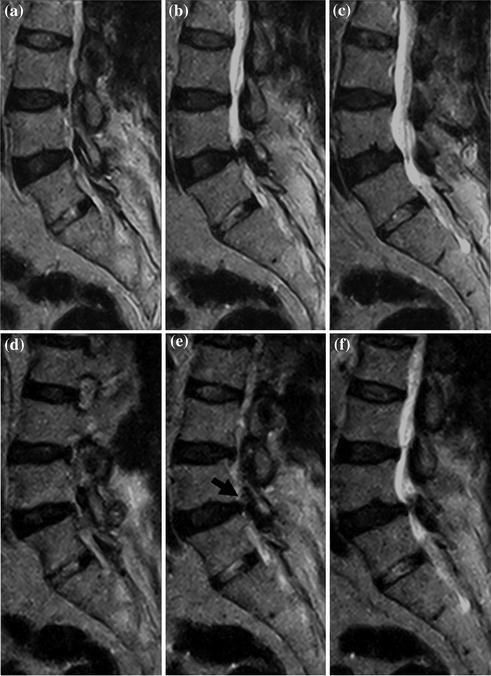
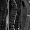
Fig. 2
a–f. FSE T2 sagittal sections in supine (a–c) and orthostatic position (d–f). Moving from supine (a–c) to orthostatic position (d–f) there is narrowing of vertebral canal at L4-L5 with incremented “impingement” of synovial cyst (arrow)
< div class='tao-gold-member'>
Only gold members can continue reading. Log In or Register to continue
Related posts:
Herniated Lumbar Disk Diskectomy
Dorsal Herniated Disk Diskectomy and Stabilization
Lumbar Stenosis and Degenerative Instability Posterior Rigid Stabilization
Cervical Spondylodiscitis Corpectomy
Traumatic Cervical Fracture-Dislocation. Conservative Treatment. Delayed Impaired Consolidation
Dorsal Collapse in Multiple Myeloma Vertebroplasty
Stay updated, free articles. Join our Telegram channel
Full access? Get Clinical Tree
