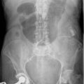
28 System for interpretation of the AXR
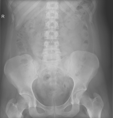
Technical factors
Also bear in mind that detail on the AXR in obese patients may be suboptimal.
Organs that can be seen
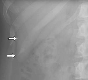
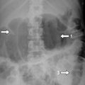
Fig. 28.2 The liver edge is subtle but can be seen (arrows). Look for liver enlargement.
Look in the region of the liver for abnormal gas in the biliary tree (see Chapter 31) or portal venous gas (see Chapter 37), which can be a poor prognostic sign in various conditions.
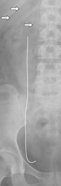
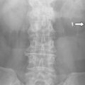
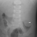
Fig. 28.3 The renal outline can also be seen, usually just lateral to the upper psoas shadow. It can be seen because it is surrounded by fat in the perinephric space (arrows). The renal outline is important for a number of reasons. Kidney stones may be seen projected over the kidney. The kidney may also be enlarged, as in polycystic kidney disease. Look for gas in emphysematous pyelonephritis (Chapter 40).
Ultrasound is the best initial modality for investigating renal disease.
Stay updated, free articles. Join our Telegram channel

Full access? Get Clinical Tree


