, Joon Woo Lee1 and Eugene Lee2
(1)
Department of Radiology, Seoul National University College of Medicine, Seoul National University Bundang Hospital, Seongnam, South Korea
(2)
Department of Radiology, Seoul National University Bundang Hospital, Seongnam, South Korea
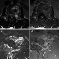
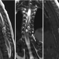
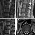
3.1 Intraosseous Tumors
Intraosseous tumors present as osteolytic or osteoblastic lesions on plain X-ray or CT images. On MRI, intraosseous tumors are best detected on T1-weighted or fat-suppressed T2-weighted images. Diffuse tumor marrow infiltration and involvement (such as may be seen in multiple myeloma) can be missed because of its ill-defined nature. The clue pointing toward diffuse bone marrow involvement is the signal of the bone marrow on T1-weighted images compared with the intervertebral discs; if the signal of the bone marrow is hypointense to intervertebral discs, diffuse marrow infiltrative tumors have to be considered.
In analyzing tumors to arrive at logical differential diagnoses, clinical factors such as patient age and tumor incidence are considered, following which tumor location and imaging pattern are evaluated.
3.1.1 Incidence-Based Approach for Intraosseous Tumors
Most common benign bone tumor | Hemangioma |
Most common malignant bone tumor | Metastasis |
3.1.2 Age-Based Approach for Intraosseous Tumors
Pediatric (< 20 years) | Eosinophilic granuloma (EG), osteoid osteoma, osteoblastoma, Ewing’s sarcoma, leukemia |
Young adult (20–39 years) | Giant cell tumor, osteosarcoma, osteoblastoma, lymphoma |
Middle age (40–59 years) | Metastasis, plasmacytoma |
Elderly (> 60 years) | Metastasis, multiple myeloma, chondrosarcoma |
3.1.3 Location-Based Approach for Intraosseous Tumors
Vertebral body | Giant cell tumor, hemangioma, benign notochordal cell tumors (BNCT), Langerhans cell histiocytosis |
Posterior elements | Sarcoma, osteoid osteoma, osteoblastoma |
Diffuse | Metastasis, multiple myeloma, lymphoma, leukemia |
3.1.4 Imaging Pattern-Based Approach for IntraosseousTumors
Benign appearing osseous lesions | Hemangioma, BNCT, EG, fibrous dysplasia |
Radiodense tumor on X-ray | Blastic metastases, osteosarcoma, fibrous dysplasia, BNCT, Hodgkin’s lymphoma, multiple myeloma |
Multi-compartmental lesions
Related posts:Stay updated, free articles. Join our Telegram channel
Full access? Get Clinical Tree
 Get Clinical Tree app for offline access

|