Randy Ray RichardsonAtlas of Pediatric Cardiac CTA2013Congenital Heart Disease10.1007/978-1-4614-0088-2_4© Springer Science+Business Media New York 2013
4. Systematic Evaluation of Cardiac CTA
(1)
Department of Radiology, St. Joseph’s Hospital and Medical Center, Creighton University School of Medicine, West Thomas Rd 350, 85013 Phoenix, AZ, USA
(2)
Children’s Heart Center, Phoenix Children’s Hospital, Phoenix, AZ, USA
Abstract
Cardiac CT evaluation requires a systematic approach to evaluate the complex anatomy in congenital heart patients. We suggest the following outline to assess these patients:
Keywords
Systematic reviewlungs and airwayscoronary arteriesgreat vesselsatriaventriclesCardiac CT evaluation requires a systematic approach to evaluate the complex anatomy in congenital heart patients. We suggest the following outline to assess these patients:
1.
Get Clinical Tree app for offline access

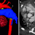
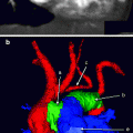
Systemic and pulmonary veins, atria, and atrioventricular (AV) valves. Start by evaluating the atria and AV valves of the heart and the veins returning blood to the heart. Look for the following:
(a)
A right superior vena cava (SVC) and inferior vena cava (IVC)
(b)
A left SVC. Check to determine whether this is solitary or if there is a bilateral SVC with or without a crossing brachiocephalic vein. Also, determine the atrial connection of the SVC; check whether it connects directly to the atrium, or if there is a connection into a coronary sinus.
(c)
Atrial connection of the IVC. Rarely, it may be bilateral or cross the spine to connect with the contralateral atrium.
(d)
Pulmonary venous return. Pulmonary veins may return blood anomalously to the systemic circulation, such as the IVC, SVC, or brachiocephalic vein. Anomalous pulmonary venous return may be partial or total depending on whether all or part of the blood goes to the systemic circulation. It also may be mixed anomalous drainage. Check for a detailed connection of each pulmonary vein.
(e)
Defects in the atrial septum. Types of defects include secundum, primum, patent foramen ovale, and sinus venosus defect. These may be difficult to see and may require viewing different phases of the scan to optimize visualization. It is helpful to correlate these images with echocardiographic examinations.
(f)
Relative size and appearance of the atria and atrial appendages.
(g)


AV valve anomalies, including atresia, stenosis, dysplasia (a dysplastic septal leaflet of the tricuspid valve is seen with Ebstein’s anomaly), a common AV valve, and thickening of the valves. Valve sizes should be provided. Subtle vegetations may be seen. Correlation with echocardiography may be helpful when subtle findings are suspected.
Related posts:
Stay updated, free articles. Join our Telegram channel
Full access? Get Clinical Tree