and Clarisse Dromain2
(1)
Department of Radiology, San Giovanni Hospital, Roma, Italy
(2)
Department of Radiology, Institut de Cancerologie Gustav Roussy, VilleJuif – Paris, France
Abstract
Taenia also called tapeworm is a parasite (cyclophillidean cestode); it infects pigs and humans and pigs are intermediate host.
Taenia
Taenia also called tapeworm is a parasite (cyclophillidean cestode); it infects pigs and humans and pigs are intermediate host.
Taenia infection is caused by ingestion of eggs shed in the feces of a human tapeworm carrier. Adult tapeworms develop (up to 2–7 m in length and produce less than 1,000 proglottids, each with approximately 50,000 eggs) and reside in the small intestine for years. Small bowel exams (X-ray follow-through and MR enterography) are able to detect a filling defect into the small bowel lumen. However diagnosis is made possible detecting proglottids or eggs in the stool.
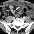
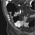
Tailgut Cyst
Tailgut cyst or retrorectal cystic hamartoma is a congenital cyst due to incomplete regression of embryonic tailgut (distal portion of the gut). It appears in imaging as a thin-walled multicystic or unilocular cyst in the presacral space adhering to sacrum, rectum, or both with extension into the ischiorectal fossa.
Tailgut cyst may be asymptomatic or lead to rectal pain, rectal bleeding, or urinary frequency. Average age at diagnosis is 35 years, and it is more commonly reported in females.
Differential diagnosis should consider perirectal abscess with anorectal fistula and mucinous adenocarcinoma and can be performed on cytological or histological basis. At drainage mucoid fluid is present and not pus.
MRI is the imaging modality of choice using phased-array coil, TSE T2-weighted sequences and contrast-enhanced T1-weighted fat saturation allowing accurate anatomical details and assessment of cyst wall in order to rule out enhancing mural nodularity/septa that may suggest the diagnosis of mucinous adenocarcinoma.
At histology tailgut cyst shows several types of epithelia plus smooth muscle.
Target Sign
Target sign is referred as a stratified appearance, a bowel segment, or a stratified enhancement pattern of bowel wall detectable at cross-sectional imaging when image is acquired orthogonal to bowel loop so that its thickened bowel wall appears as multiple concentric rings with different attenuation/signal intensity or different degrees of contrast enhancement.
Target sign was firstly described by Balthazar in 1991 using CT imaging referring to a contrast-enhancement pattern of a symmetric thickened bowel wall presenting three layers (inner enhancing layer, outer enhancing layer, intermediate layer with reduced attenuation with respect to the enhancing layers). The explanation of these findings regards different degrees of inflammation and edema of the bowel wall with hyperemia of the mucosa (inner enhancing layer) and serosa/muscularis propria (outer layer) with edema of submucosa (intermediate non-enhancing layer). The target sign is best appreciable on arterial and portal venous phase; in case of marked submucosa edema, it can be detected also at non-enhanced study. The same enhancement pattern is detectable also at MRI with the same meaning. Inner low attenuation layer may also represent fatty deposit into submucosa layer (check quantitatively HU at non-enhanced CT or analyze SI on T1-weighted and fat-suppressed T1-weighted MR images) and is associated to chronic inflammation (proctitis after radiation therapy or ulcerative colitis or Crohn’s disease).
While nonspecific the target sign may allow to suggest the inflammatory origin of bowel wall thickening.
Target sign is described in several conditions, most of them addressed as inflammatory changes of bowel wall (appendicitis, CMV colitis, Crohn’s disease, Henoch–Schonlein purpura, ischemic enteritis or colitis, pseudomembranous colitis, radiation-induced enteritis, ulcerative colitis, and bowel edema associated with portal hypertension). However also intussusception showed a target appearance due to the concentric “telescopic” configuration of intussuscipiens and intussusceptum bowel wall and mesenteric fat.
TEM
Transanal endoscopic microsurgery (TEM) is a minimally invasive surgical technique, which is performed endoluminally and is optically enhanced (usually through stereoscopic vision). This is an organ preservation technique with local excision (disc excision) of the rectal wall without lymph node removal that allows the patient to recover rapidly. The whole procedure is performed transanally.
< div class='tao-gold-member'>
Only gold members can continue reading. Log In or Register to continue

Stay updated, free articles. Join our Telegram channel

Full access? Get Clinical Tree


