Fig. 15.1
Pelvic tilt after hip injection or hyaluronic acid
Fig. 15.2
Exercise of Leg Extention
Fig. 15.3
Strengthening of the gluteal muscles
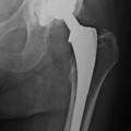
15.2 Rehabilitation After Hip Prosthesis
The hip is the proximal joint of the inferior limb. While being an enarthrosis from the anatomical point of view, it has three degrees of freedom:
1.
Transversal–along which the movement of extension flex is effected.
2.
Antero-posterior–along which the movements of abduction and adduction are effected
Hip osteoarthritis is one of the most serious diseases of the motor organs, most commonly associated with aging. It leads to loss of independence and worsening of disability in elderly individuals. The disease causes pain which results in gait disorders. Hip osteoarthritis is prevalent in all races and is statistically more common in women than in men. According to latest literature reports, the overall incidence is estimated at 2–3 % of global population [8]. Total hip replacements are performed most commonly because of progressively worsening of severe arthritis in the hip joint. The most common type of arthritis leading to total hip replacement is degenerative osteoarthritis of the hip joint. This type of arthritis is generally seen with aging, congenital abnormality of the hip joint, or prior trauma to the hip joint. Other conditions leading to total hip replacement include bony fractures of the hip joint, rheumatoid arthritis, and septic necrosis, or a vascular necrosis, of the hip bone. Certainly, another frequent cause is hip fracture often due to osteoporosis. The progressively intense chronic pain together with impairment of daily function including walking, climbing stairs, and even arising from a sitting position, eventually become reasons to consider a total hip replacement. Because replaced hip joints can fail with time, whether and when to perform total hip replacement are not easy decisions, especially in younger patients. Replacement is generally considered when pain becomes so severe that it impedes normal function despite an intensive medical and rehabilitation protocol. An important way to evaluate the disability and the pain in those who undergo a surgical joint replacement is to use a validated evaluation scale. Every patient should be evaluated by the administration of these useful tools.
One of these evaluation scales is the Western Ontario and McMaster Universities Arthritis Index (WOMAC), that is widely used by health professionals to evaluate the condition of patients with osteoarthritis of the knee and hip and includes the evaluation of pain, stiffness, and physical functioning of the joints. The WOMAC measures five items for pain (score range 0–20), two for stiffness (score range 0–8), and 17 for functional limitation (score range 0–68). Physical functioning questions cover everyday activities such as stair use, standing up from a sitting or lying position, standing, bending, walking, getting in and out of a car, shopping, putting on or taking off socks, lying in bed, getting in or out of a bath, sitting, and heavy and light household duties [9, 10].
Another useful questionnaire is the Harris hip Score (HHS) that can be used before and after the hip joint replacement in order to evaluate the pain and the disability after the surgical intervention and after the rehabilitation program. Some studies about the utility of using the HHS were conducted by a group of researchers from the Department of Orthopedics and Traumatology of the Medical University in Ismir, Turkey. One of these studies was conducted on a group of 60 patients subjected to total hip replacement and divided into two groups. Patients in the study group received preoperative physical therapy to improve the muscle strength in upper and lower limbs as well as the range of motion of the hip joint starting from 8 weeks before the surgery. The group also participated in rehabilitation programs regarding living with endoprosthesis. The control group received neither physical therapy nor education. The patients were evaluated in a physical assessment immediately before the procedure, at discharge, at 3 months and 2 years after the surgery. HHS, visual analog scale, and the hip abduction range of motion were evaluated. Although patients in the study group were activated earlier than the patients in the control group, there were no differences between both groups at hospital discharge. The respective p values were<0.48 for the HHS,<0.97 for the hip abduction range of motion,<0.54 for the visual analog scale, and<0.89 for the activity. As regards further follow-up, both groups showed improvement in the HHS 2 years after the surgery [11].
Another useful scale is the Short Form 36 (SF-36). The SF-36 is a 36-item questionnaire that produces scores in eight domains relating to the patient’s quality of life. These are physical functioning, role limitation due to physical problems, bodily pain, general health perception, emotional vitality, social functioning, role limitation due to emotional problems, and mental health [12]. Another important evaluation scale is the FIM (functional independence measure) that analyzes the assistance and involves that the patient complete a questionnaire on the activities of daily living (ADLs) and comprises two groups of functions: cognitive (communication and relational/cognitive capacity) and motor (care of the sphincter of the person, mobility, locomotion and control) [13]. Another evaluation scale is the Barthel index. The Barthel scale or Barthel ADL index is an ordinal scale used to measure performance in ADL. Each performance item is rated on this scale with a given number of points assigned to each level or ranking. It uses ten variables describing ADL and mobility. A higher number is associated with a greater likelihood of being able to live at home with a degree of independence following discharge from hospital. The amount of time and physical assistance required to perform each item are used in determining the assigned value of each item. External factors within the environment affect the score of each item. The scale was introduced in 1965 and yielded a score of 0–20. Although this original version is still widely used, it was modified by Granger et al. in 1979, when it came to include 0–10 points for every variable, and further refinements were introduced in 1989. The Barthel index designed at the original scale was insensitive to change and had arbitrary scores. The sensitized version sharply discriminates between good and better and poor and poorer performances. The Barthel index has been shown to have portability and has been used in 16 major diagnostic conditions. It has demonstrated high interrator reliability (0.95) and test-retest reliability (0.89) as well as high correlations (0.74–0.8) with other measures of physical disability [14].
Prior to surgery, there is a general deficit in muscular strength along the affected limb as compared to the contralateral (healthy) side in patients with unilateral hip osteoarthritis (OA), and muscles such as the abductors, vastii, rectus femurs, and psoas show marked atrophy. This is evidenced by reduced cross-sectional area and infiltration of noncontractile components, i.e., an average 10 % increase in fatty infiltration of muscle (myosteatosis) in the affected limb compared to the healthy one. As well as reducing muscle strength, myosteatosis also exacerbates fall risk. This muscular dysfunction is likely to contribute to the reduced walking ability of OA patients, as loss of lower limb muscle strength has been shown to predict the onset of activities of daily living dependence in the elderly.
Consistent with the persisting functional deficits following surgery, these atrophic changes about the hip are still evident up to 2 years following total hip replacement (THR) surgery. Frail elderly persons with sarcopenia often undergo musculoskeletal-related surgery such as THR, and the hospitalization-associated immobilization further compromises the skeletal system, with potentially grave consequences. Earlier operation may prevent the development of persistent atrophic changes that occur after THR and there is a suggestion that fatty infiltration may be reversed by intensive rehabilitation. There have been considerable technical efforts toward optimizing surgical treatment of patients with arthritis of the hip, for example with over 100 varieties of hip prostheses being available, multiple types of bearing couples and several surgical approaches. As technology and surgical techniques for THR improve, patient expectations, including for an early return to normal physical function and activities, have also increased. However, the actual health gain for many of these innovations relative to “standard” THR is small in terms of patient function and quality of life. In the past, a prolonged hospital stay after THR surgery incorporated a period of supervised rehabilitation to try to achieve restoration of physical function. However, due to the introduction of initiatives such as integrated care pathways and considerations of cost and increasing patient satisfaction, the length of hospital stay following joint replacement has been substantially reduced. Standard physiotherapy (i.e., not involving resistance training) following major surgery enables most patients to regain basal levels of function but leaves them with significant muscle wasting as it lacks the intensity of exercise required to elicit muscle hypertrophy.
The most commonly used rehabilitation regimes for elderly individuals are based on functional types of exercises without external loading. However, this type of intervention not only fails to elicit increases in muscle mass but does not prevent further muscle atrophy. In contrast, high-intensity PRT is an extremely effective and safe method for inducing muscle hypertrophy and increasing muscle strength and subsequently improving functional performance in healthy individuals, those with chronic disease, e.g., rheumatoid arthritis, and the elderly. THR surgery provides good relief for patients’ pain but fails to fully restore physical function. A systematic review demonstrates that significant improvements in muscle strength and function are achievable with PRT. Regardless of the timing of the intervention, PRT appears to have a significant benefit on patient function following THR. Late PRT interventions do work and are safe, and they have been performed mainly in the home setting, but the studies done have short periods of follow-up and have a further limitation of the preexisting functional deficit due to the timing postoperatively. Early PRT regimes identified in the studies reviewed in this article have shown the need for a center-based approach, and this has demonstrable benefit, but there are issues of high costs of transport and supervision. Early home-based PRT studies that are effective and safe with adequate follow-up after THR surgery would potentially address these issues [15].
Also, the rehabilitative treatment before the surgical intervention can have utility in order to prepare the patient to the postoperational phase. It is important to educate the patient on the correct use of the aids and to precautions to be taken. Another remarkable aspect can be that of the respiratory reeducation that, besides allowing muscular relaxation, it allows to patients to work on improving their respiratory performance. It is important also to improve the ventilation of the pulmonary base functions associated with postural drainage and to apply maneuvers of vibration and of clapping. To prevent pulmonary stasis, the subject must perform exercises involving the abdominal muscle, the diaphragmatic dome, and intercostal muscles [16].
After total hip joint replacement surgery, patients must immediately start the rehabilitation program. On the first day after surgery, it is common to begin with some minor physical therapy while sitting in a chair. Initially, supportive devices such as a walker or crutches are used. Pain is monitored while exercises take place. Specific techniques of body posturing, sitting, and using an elevated toilet seat can be extremely helpful. Patients are instructed not to cross the operated lower extremity across the midline of the body (not crossing the leg over the other leg) because of the risk of dislocating the replaced joint. They are discouraged from bending at the waist and are instructed to use a pillow between the legs when lying on the nonoperated side in order to prevent the operated lower extremity from crossing over the midline. Another important attention is to keep the operated leg always extended during the sitting position in order to limit the hip flexion. In fact, with the lower arm completely extended, it is physiologically impossible to flex the hip more than 90° [7]. The incidence of luxation decreases more than 95 % after 12 week from the surgical intervention. When the scar is completely closed, it is important to work manually on it in order to make it more elastic and less adherent to the tissues below. Patients are given home exercise programs to strengthen the muscles around the buttocks and thigh. Most patients attend outpatient physical therapy for a period of time while incorporating home exercises regularly into their daily life. Patients will continue to use supportive devices as monitored and recommended by the attending physician. Gradually, patients become more confident and less dependent on supportive devices. Patient education is important to ensure longevity of the replaced hip. Strenuous exercises such as running are discouraged. The best way to improve muscle strength and promoting mobility and endurance is to perform a period of hydrokinesitherapy. Unfortunately, it is not easy to find a place in where to perform hydrokinesitherapy, remembering that the physiotherapist needs to become further specialized in order to be able to follow the patient in water. In a second phase, when the supportive devices are dismissed, one can send the patient to a swimming pool to improve muscle strength and to promote mobility and endurance. The work in the swimming pool is important to favor a complete muscular relaxation stimulating the aerobic general capacities and to limit the risk of traumas from fall. The exercises in water allow the possibility to effect a work to variable load. The action of the water also allows a vascular exercise that normalizes the speed of the blood flux by allowing more valid metabolic exchanges. The therapeutic exercise in heated swimming pools acts across three fundamental principles: the hydrostatic effect based on the principle of Archimedes that allows floating and stimulates proprioception; the hydrodynamic effect that is represented by the resistance of the liquid and that allows the execution of exercises against resistance; and the thermal effect that induces muscular relaxation and sedative action reducing the reactions of defense [16].
The rehabilitative program can be that shown from Table 15.11
Table 15.1
Rehabilitative program
Days | Program |
|---|---|
Day of the intervention | Exercises of breathing; exercises of ROM of the ankle |
1st day after the surgery | Isometric exercises of the quadriceps, of the gluteal muscle (Valuate the surgical intervention), maintain the buttock in abduction, active-helped mobilization, exercises of bending of the knees |
2nd, 6th day after the surgery | Weight bearing as tolerated. Deambulation with the use of a walker device or crutches |
7th day—3rd month after the surgery |
15.3 Rehabilitation After Knee and Ankle Prosthesis
15.3.1 Knee Joint Prosthesis
The knee joint has three degrees of freedom. The flex extension is the principal movement; rotation and traverse movement are realizable only with flexed knees. This is important in order to guarantee the double requirement of the stability in extension to sustain the load and of the mobility in flexion to adapt the gait to irregularities of the ground [7]. Knee prostheses are now surgically applied with good results. Clinical data show that the greater the functional deterioration of the range of motion before the operation, the slower the recovery and rehabilitation after the surgical intervention. Furthermore, the patient may have activated a strategy of compensation with alterations at the level of the pelvis, trunk, and inferior limbs. The unbearable pain associated with serious functional limitations is the principal indication for surgical intervention. It is fundamental to use appropriate evaluation scales, such as:
Rivermead Mobility Index (RMI)–a measure of disability related to bodily mobility. It demonstrates the patient’s ability to move her or his own body. It does not measure the effective use of a wheelchair or the mobility when aided by someone else [18].
Barthel index (BI)–an ordinal scale used to measure performance in activities of daily living (ADLs) as described before.
Knee Clinical Society System rating–an easily reproducible evaluation scale that measures the capacities of the subject (deambulation, climb, descending stairs, use of aids) and also the articular function of the knees (pain, degree of movement, muscular forces, articular stability, alignment, contracture in bending, and deficit of extension) [19].
Functional independences measure (FIM)–analyzes the assistance needed by the patient by completing a questionnaire on activities of daily living (ADLs). It considers two groups of functions: cognitive (communication and relational/cognitive capacity) and motor (care of the sphincter, mobility, locomotion, and control) [13].
Certainly, in these circumstances it is important to have prior rehabilitative preparation of the surgical intervention. The result of the surgical intervention depends on a careful clinical evaluation of the joint but also requires a change of certain unfavorable parameters: overweight, general state of the musculoskeletal and cardiovascular systems. In fact, all of these features may be provoked by the so-called hypokinetic syndrome, a condition of reduced mobility due to pain, which considerably limits their mobility. The evaluation of the range of motion and of muscle status is of paramount importance (Fig. 15.4) [20]. Also important is a program of exercises of respiratory physiotherapy to improve bronchial drainage. Before prescribing the physiotherapy, it is important to be informed about the sort of surgical intervention and the sort of positioned prosthesis. The motives for applying total prosthesis versus partial ones depends on various factors. The functional load is often not equally distributed and the damage may be limited to only some parts of the knee. The rehabilitative treatment must be aimed at restoring maximum safety, autonomy of walking, and best possible activities of daily life (ADL).
The objective in the first phase is to alleviate surgical stress, to fastly recover the mobility of the knees, and to control the maintenance of the normal articular mechanics. Right after the surgical intervention it is important to control the pain that causes muscular reflex inhibition. Antithrombosis elastic stockings should be worn. The external compression allows the reduction in the circumference of the limb and increases blood flux, decreasing venous stasis and the risk of the formation of thrombosis. Elastic stockings should effect decreasing compression by the ankle towards the thigh by increasing the blood flux in femoral veins. The mobilization of the operated limb must begin early on: passive continuous movement allows to recover the articular mobility and to prevent functional limitations [21]. One can make a plan of the exact parameters of time and range of passive continuous movement. The duration of the sessions must be progressively increased and must be carried several hours per day. The speed must be constant and low in order to avoid inflammatory reactions. The patient must be consciously aware of automatic movements to prevent defense reactions or unintentional movements. All this is aimed at rapidly attaining a 90-degree knee flexion. Another important issue is the mobilization of the patella, which prevents adhesion to the surrounding tissues (Fig. 15.5).
Proprioceptive excercises are also necessary for recovering coordination (Fig. 15.6). At this point, an electromyographic evaluation may be advisable to determine possible differences of force between agonists and antagonists. It may also be useful to carry out specific protocols of electromyographic biofeedback. These tools allow an early recovery of the muscle tone involving the patient directly in active and motivating excercises [22]. The role of the physiotherapist is not limited to improving the muscle tone and coordination, but also to preventing antalgic contractures that can limit rehabilitation and loosen the joint. Exercises dedicated to the hamstrings muscles are also important. The cyclette used at low resistance is an useful exercise at this stage to improve the range of motions; it must be adjusted to ‘high saddle’ [16].
The best way to improve muscle strength and promoting mobility and endurance is to schedule hydrokinesitherapy, as described above. Also important is the correction of possible differences in length of the inferior limbs to balance and to stabilize deambulation.
Fig. 15.4
Evaluation of the knee joint and muscle of the leg
Fig. 15.5
Mobilization lower limb
Fig. 15.6
Proprioceptive exercises for the lower limb
15.3.2 Ankle Joint Prosthesis
Surgery of painful, end-stage ankle arthritis includes ankle arthrodesis and total ankle replacement. In the past decade, total ankle replacement has become a viable alternative to ankle arthrodesis. Modern implant designs either involve a syndesmosis fusion and resurfacing of the medial and lateral recesses of the ankle joint or the use of a three-component, mobile-bearing implant. In limited clinical series, early results of both of these prosthetic design approaches have been encouraging. In selected patients, ankle arthroplasty is an effective approach to relieving pain and improving function [23].
Ankle arthrodesis has been the treatment of choice for patients who have arthritic or degenerative ankle disease with disabling pain and loss of motion. But the results of long-term retrospective studies show that a significant percentage of arthrodeses have been unacceptable to patients and are considered to be clinically poor with many failures by the physicians. Therefore, total replacement of the ankle joint is becoming recognized as an alternative to ankle arthrodesis. This procedure eliminates pain and provides sufficient ankle motion for resumption of normal activities. Indications and contraindications for total ankle replacement are analogous to those for hip and knee joint replacement. Surgery is considered only when conservative treatment has been attempted with no improvement. Total ankle joint replacement is indicated when rheumatoid arthritis or osteoarthritis causes advanced arthritic changes with disabling pain and loss of ankle motion. Traumatic degenerative disease following ankle fracture, recurrent sprains, and injuries to the talus that could result in avascular necrosis or osteochondritis are also indications. In certain cases, total ankle joint replacement is also indicated following unsuccessful ankle arthrodesis. The rehabilitation protocols that are used actually are founded on not very recent work or are based on empirical observations.
Before surgery, the physiatrist counsels the patient regarding the procedure and explains the treatment program and goals. The patient’s range of motion and muscle tone is evaluated, with special attention to the involved extremity (Fig. 15.7). The patient’s ability in ADLs is assessed. The gait pattern of the patient is analyzed and gait deviations are recorded. All of the findings are documented. The patient is instructed in deep breathing and coughing exercises. Bilateral isometric contractions are taught for the gluteus maximus and quadriceps femur muscles. Isotonic ankle exercises for plantar flexion and dorsiflexion are taught for the uninvolved extremity. After surgery, the entire foot is wrapped in a bulky pressure dressing and elevated until the swelling has diminished and the joint feels comfortable to the patient. Generally, this requires three to five days [24].
Fig. 15.7
Ankle mobilization
Related posts:

Stay updated, free articles. Join our Telegram channel

Full access? Get Clinical Tree


