KEY FACTS
Terminology
- •
Infiltrative neoplasm of testis in which tumor cells surround and compress seminiferous tubules and normal testicular vessels
Imaging
- •
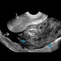
Bilateral, solid, hypoechoic, hypervascular nodules/masses
- •
Diffuse hypoechoic testis with hypervascularity
- •
Striated pattern
- •
Testicular shape not altered
- •
Normal testicular vessels with straight course crossing through lesions
Pathology
- •
Most commonly secondary lymphomatous involvement of testis; rarely primary
- •
Lymphoma behaves similar to leukemia with abnormal cells diffusely infiltrating interstitium with compression of seminiferous tubules without causing their destruction
- •
Testis is “sanctuary organ”: Blood gonad barrier limits accumulation of chemotherapeutic agents
Clinical Issues
- •
Stages IE and IIE: Orchidectomy
- •
Stages IIIE and IVE: Systemic chemotherapy using cyclophosphamide, doxorubicin, vincristine, and prednisolone
- •
Radiation in symptomatic and bulky deposits
- •
Lymphoma accounts for ~ 5% of all testicular tumors
- •
Most common bilateral testicular tumor
Scanning Tips
- •
Side-by-side comparison of both testes in single image with both grayscale and color Doppler is essential to assess for symmetry in size, echogenicity, and vascularity
- ○
Do not use dual image/split screen because scan parameters, such as TGC or color Doppler scale, may be altered between windows
- ○