Testicular Torsion
Parth C. Patel
Cassandra M. Sams
CLINICAL HISTORY
16-year-old male with a history of acute onset of scrotal pain.
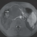
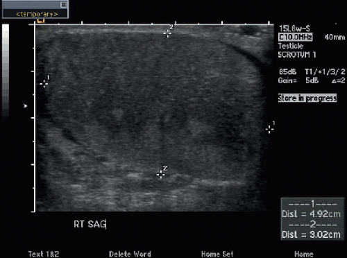 FIGURE 31A |
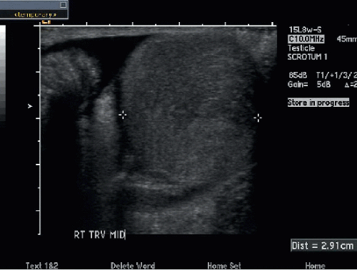 FIGURE 31B |
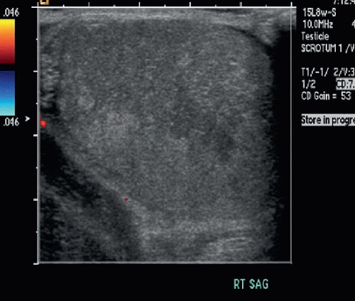 FIGURE 31C |
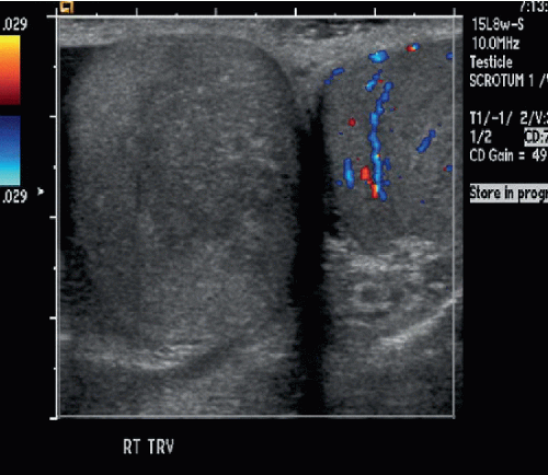 FIGURE 31D |
FINDINGS
Figures 31A and 31B: Longitudinal and transverse grayscale US images show heterogeneous echotexture of the abnormal right testicle (between cursors). Figure 31C: Color Doppler sonogram of the right testicle shows absence of blood flow. Figure 31D: Color Doppler image of the right and left testicles on the same image for comparison demonstrates absence of flow in the right testicle and flow in the left testicle. At surgery, bilateral bell-clapper deformities were identified. The right testicle was detorsed and viable. Bilateral orchiopexies were performed.
DIFFERENTIAL DIAGNOSIS
Epididymo-orchitis, testicular torsion, torsion of the appendix testis, testicular abscess, testicular tumor, testicular trauma.
DIAGNOSIS
Related posts:


Stay updated, free articles. Join our Telegram channel

Full access? Get Clinical Tree


