CHAPTER 6 The abnormal hilum
6.1 Unilateral hilar enlargement
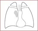
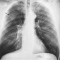
Hilar enlargement is difficult! Suspect unilateral hilar enlargement if:
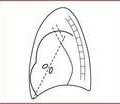
3. There is a loss of the normal concave shape – the hila are usually concave in shape. This concavity may disappear and be the first sign of hilar enlargement.
If you suspect unilateral hilar enlargement then:
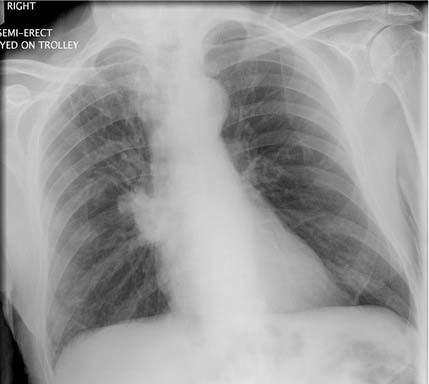
1. Check the technical quality of the film. A rotated film will make one hilum appear larger than another.
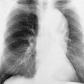
2. Look at the lateral film. An enlarged hilum may look abnormally dense on the lateral and sometimes this is easier to spot than on the PA.
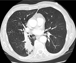
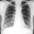
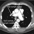
Now you need to decide whether the enlargement is due to enlarged vascular shadows or enlargement of the hilar lymph nodes or whether it is due to a central bronchial carcinoma superimposed over the hilar shadow. These are the likely possibilities.
Stay updated, free articles. Join our Telegram channel

Full access? Get Clinical Tree


