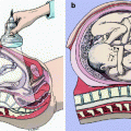
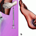
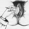
Fig. 7.1
Vaginal digital examination in the assessment of fetal head station. Fetal head is defined as engaged when its most leading point is at or below the imaginary line passing through the ischial spines. The different stations are 1 cm above (negative sign) and below (positive sign) this imaginary line
The most important factor not taken in consideration with this assessment is the peculiar shape of the pelvic axis, the so-called Carus curve [2]. The pathway of the fetal head through the pelvis is not a straight line but a curvilinear one. More specifically the birth canal can be divided in two sections. In the upper section (clinical station −5 to 0), the fetal head travels along a straight line until it reaches the reference point at station 0 (Fig. 7.2, panel a). From here to delivery, the fetal head traverses a path that is best represented by a curved line (clinical station +1 to +5) (Fig. 7.2, panel b). In fact, delivery occurs by extension of the previously flexed engaged head (Fig. 7.2, panel c). The occiput descend until it contacts the end of the pubic symphysis, which acts as a fulcrum around which the fetal head changes direction, extending through the last curvilinear portion of the birth canal (Fig. 7.2, panel d). From this fundamental knowledge of physiology comes the need of finding a more reliable and objective tool to correctly assess fetal head station and descent in the birth canal. This is of course of extreme importance in deciding whether or not to attempt a vaginal delivery either spontaneous or operative, or to perform a cesarean section.
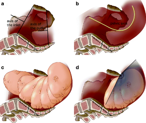
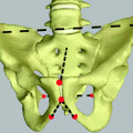
Fig. 7.2
Pelvic axis that fetal head needs to follow in its progression in the birth canal. Panel a: Two different axis are present, one in the upper portion and one in the lower portion. Panel b: The pelvic axis is better represented by a curved line (Carus curve) [2]. Panel c: Delivery occurs by extension of the previously flexed engaged head. Panel d: The lower margin of the symphysis acts as a fulcrum around which fetal head extends
7.2 Use of Transperineal Ultrasound
Since 1977, ultrasound has been reported as an adjunctive method for evaluating the level of the fetal head within the maternal pelvis.
Lewin et al. [3] positioned an ultrasound probe on the mother’s skin at the level of the sacral tip oriented toward her umbilicus. In this way, they were able to record the distance between the sacrum and the fetal head circumference. Their observations though were confined to the upper straight portion of the birth canal, above the Carus curve.
Richey et al. [4] evaluated head station utilizing the linear measurement drawn from the perineum to the fetal head. Their correlation between measurement of the station of the fetal presenting part by transperineal ultrasound and vaginal digital examination (VDE) were once again limited to the stations above the ischial spines.
Sherer and Abulafia [5] showed an 85.6% agreement between transvaginal digital and transabdominal ultrasound determination of fetal head engagement. Their evaluation, however, was limited only to fetal head engagement.
Dietz and Lanzarone [6] used the pubic symphysis and the most distal point of fetal head contour as a landmark in quantifying head engagement using translabial ultrasound imaging. Their method showed a reasonable correlation with fetal station assessed digitally, but the study was conducted in patients whose fetal station was not lower than +1. Although the technique seemed to be useful in high stations, at which the curved nature of the lower portion of the birth canal is not relevant, it was not tested in those patients for whom it is most important to accurately estimate station, those with fetal head station below +1.
Heinrich et al. [7] reported their experience with a translabial ultrasound approach. He was able to identify the pubic symphysis joint, the widest diameter of the fetal skull, the “infrapubic line” running perpendicular to the long axis of the symphysis and originating from its caudal end, and the “head direction” defined as the direction of a line perpendicular to the widest diameter of the fetal head in the infrapubic plane, with respect to the infrapubic line. In this study though the widest diameter of the fetal head was defined arbitrarily as well as its relationship with the interspinous plane.
Eggebo et al. [8] reported that a fetal head-perineum distance measured by transperineal imaging can predict vaginal delivery after induction of labor. They showed an association, but not a direct relationship, between fetal head-perineum distance and fetal head station. Unfortunately, these measurements may vary with the degree of compression of the soft tissue, affecting the real station of the fetal head in the birth canal. Furthermore, the positioning of the outer bony limit on the fetal skull was chosen arbitrarily.
Ghi et al. [9] reported their experience on the use of translabial ultrasound following fetal head descent in the birth canal reporting three different categories of fetal head direction: (1) downward direction, with the head in the upper third of the pelvis; (2) horizontal direction, with the head in the midplevis; and (3) upward direction, with the head most likely in the lower third of the pelvis. They concluded that translabial sonography allows a diagnosis of fetal station with accuracy comparable to that of digital examination.
7.3 The “Angle of Progression”
In all these studies using the transperineal/translabial ultrasound approach, none of the techniques have concentrated on the arc-like pathway that the head must follow to move through the upper and lower portions of the birth canal. No attention has been placed on monitoring the true unit that changes during the progression in the birth canal: the angle of flexion and extension of the fetal head. The fetal head will be able to engage only if it will be completely flexed with the chin touching the fetal sternum. This will allow the smallest anteroposterior diameter of the head, namely, the suboccipitobregmatic diameter, to face and progress through the inlet-, mid-, and lower pelvic diameter, respectively the oblique and the two anteroposterior. At the lower segment of the upper portion of the birth canal, the head will extend following the curve of the lower portion of the birth canal, being delivered due to the extension mechanism that will use the lower margin of the pubic symphysis as a fulcrum around which the fetal head will change direction (Fig. 7.2, panel d).
Having this anatomical concept very clear, we started assessing the feasibility and reproducibility of measuring station and observing descent of the fetal head using transperineal ultrasound (TPU) examination during labor. Another objective of our investigation was to compare the clinical evaluation of fetal station through vaginal digital examinations with concomitant TPU assessments of station. Lastly, we examined trends in TPU data in laboring patients that could help to identify those who would go on to deliver vaginally rather than needing cesarean section for failure to progress. This, in turn, will be useful in developing a decision-making strategy to determine the need for operative delivery in patients progressing slowly through the second stage of labor.
7.4 Population
Our original work started in Italy in 1996 and was completed in the USA in 2004. Eighty-eight pregnant women in labor were included in the study (47 women in Milan, Italy, and 41 women in Denver, CO, USA) [10–12]. All women had a live singleton pregnancy with the fetus in cephalic presentation and a gestational age of more than 37 completed weeks dated by first- and/or midtrimester ultrasound.
7.5 Real-Time Ultrasound Measurements
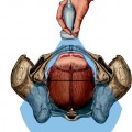
Patients were examined in their labor rooms. A 5.0-MHz ultrasound probe was enclosed in a latex glove covered with ultrasound gel and was then placed between the labia below the pubic symphysis. The sagittal view, in which the long axis of the pubic symphysis could be visualized, was obtained by gently rocking the transducer upward. While in this same plane, the fetal head could easily be discerned and its skull line identified. On the sagittal image, a line was drawn on the screen between calipers placed at the two points identifying the long axis of the pubic symphysis. A second caliper line was then created on the frozen image that extended from the most inferior portion of the pubic symphysis tangentially to the fetal skull contour. The angle between the constructed lines was then measured (Fig. 7.3, panel a–c). The symphysis pubis may have varying ultrasound appearance (Fig. 7.4), and because it is a rather short structure, care was taken to identify its exact ends so that the long axis could be precisely determined. The transducer was maintained perfectly median to avoid the acoustic shadow given by the adjacent superior pubic rami (Fig. 7.5). The use of the sagittal view also allowed clear identification of caput succedaneum (Fig. 7.6) and molding (Fig. 7.7) when present. All of the images were obtained with an empty bladder. TPU scans were performed at different times during labor for the 88 subjects included in the study.
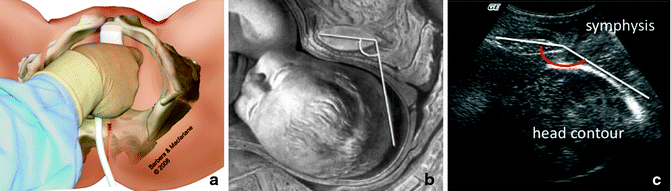
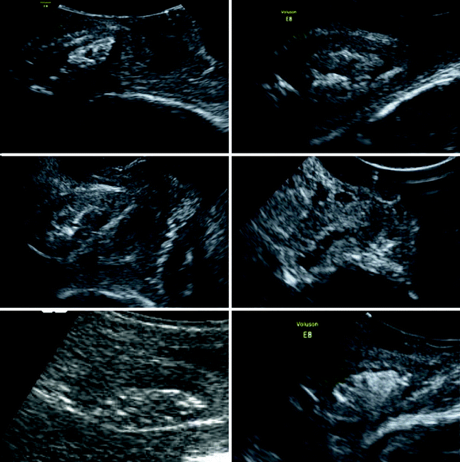
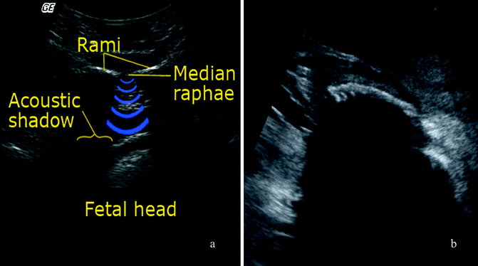
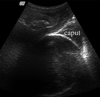
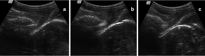
Fig. 7.3
Transperineal ultrasound (TPU) in the assessment of the angle of progression. Panel a: Practical technique for TPU, with glove covering the ultrasound probe in a sagittal section. The probe is moved gently side by side to see the gravel appearance of the symphysis in between the two rami. Panel b: Wax representation of fetal head in the birth canal and relationship between symphysis pubic and fetal head contour. Panel c: Ultrasound appearance of the measurement of the angle of progression. The upper and lower pole of the symphysis are clearly identified allowing the drawing of its long axis; a line is drawn from its inferior pole tangentially to the fetal head contour easily identified
Fig. 7.4
The symphysis pubis has most of the time a gravel appearance, sometimes hyperechoic and some other times hypoechoic, depending on its water content. It is important though to get a median scan to avoid acoustic shadow given by the adjacent superior pubic rami
Fig. 7.5
Panel a: Suprapubic transverse section showing the median raphae (symphysis pubic) and the acoustic shadow given by the 2 superior pubic rami. It is through the median raphae that the ultrasound beams will pass identifying the gravel appearance of the symphysis. Panel b: Paramedian transperineal section affected by the acoustic shadow given by the superior pubic rami (to be avoided)
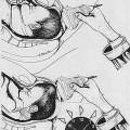
Fig. 7.6
Sagittal view of transperineal ultrasound showing how easy it is to distinguish the caput succedaneum from the fetal skull
Fig. 7.7
Sagittal view of transperineal ultrasound showing absent (a), minimal (b), and marked (c) molding
For assessments performed during the second stage of labor, the time was noted and later used to calculate the interval from scanning to delivery. In all cases, measurements were performed in concert with digital examinations. All patients tolerated the TPU procedure well, without any reported discomfort. Ultrasound assessment of the head station never took more than 1 min.
7.6 Intraobserver and Interobserver Variability
Seventy-five of the 86 patients studied had one set of between 2 and 5 replicated scans obtained at approximately the same time. These 75 women provided a total of 172 sets of scans, which were used to assess intraobserver variability.
In order to assess interobserver variability, a second independent and well-trained observer, blinded to the other’s results, obtained 15 duplicate sets of scans at distinct times of labor among 12 randomly selected women as generated by the primary observer. The consecutive scans were performed with no more than 3 min between the assessments of the two observers. The goal was to generate paired sets of replicated images under nearly identical conditions.
The intraobserver variability was calculated to be 2.96°, whereas the interobserver variability was of 1.24° [12].
7.7 Assessment of Clinical Head Station
Assessments of fetal head station were performed by vaginal digital examination (VDE) conducted by the managing clinician, who was not involved in the study and was blinded to the ultrasound data. These examiners included a senior consultant and a mixture of residents and fellows with at least 3 years of experience.
Station was determined by assessing the relationship between the most distal cranial point and the level of the ischial spines. The timing of the clinical station assessments coincided with TPU assessments during the various stages of labor so that correlations and associations between these two distinct methods could be evaluated statistically.
There was a significant linear correlation between the angle of progression measured on TPU and the clinical station assessed by digital examination (R 2 = 0.2659, P < 0.001) (Fig. 7.8).
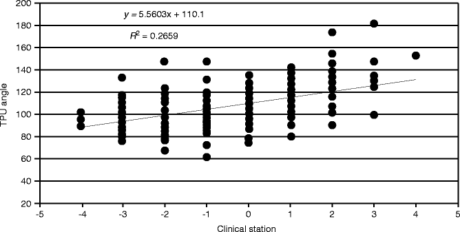
Fig. 7.8
Relationship between angle of progression measured by transperineal ultrasound and fetal head station assessed by vaginal digital examination (Barbera et al. [12], with permission)
Because of the large standard deviations for each station (roughly nine times the standard error), the prediction intervals of TPU angle seriously overlap, thereby compromising any precision in predicting TPU angles from clinical station assessed by vaginal digital examination. In turn, the clinical station as assessed by VDE can vary widely at any given TPU angle of progression, which we believe represents a more objective variable.
Related posts:
 General Intrapartum Sonography Setup and Use in Labor
 Intrapartum Sonography and Clinical Risk Management
 New Technologies for Monitoring Labor Progress
 Clinical Evaluation of Labor and Intrapartum Sonography
 Occiput Posterior Position and Intrapartum Sonography
 Intrapartum Sonography and Labor Progression
Stay updated, free articles. Join our Telegram channel

Full access? Get Clinical Tree


