Fig. 11.1
Evaluation of the lateral ankle to assess the peroneal tendons. (a) The probe and patient are positioned to evaluate the peroneal tendons on a short-axis scan. (b) Anatomical scheme of the peroneal tendons as seen along their short axis, F fibula, PL peroneus longus, PB peroneus brevis. (c) US long-axis scan of peroneal tendons
Medial Compartment
The patient lies supine on the bed, with the knee flexed at about 90°, and the foot is slightly externally rotated.
Tarsal Tunnel
The posterior tibial tendon, the flexor digitorum longus tendon, the tibial neurovascular bundle, and the flexor hallucis longus tendon are contained in the tarsal tunnel (medial to lateral).
The tarsal tunnel can be assessed on axial scans placing one edge of the probe on the tip of the medial malleolus and the other on the Achilles tendon. The posterior tibial tendon must be evaluated along its whole course with axial scans, up to its main insertion on the navicular bone. This area must be assessed carefully, also with longitudinal scans, due to the complexity of the enthesis that could result in anisotropy artifacts. The presence of an accessory navicular bone is an extremely common finding. The flexor digitorum longus and flexor hallucis longus tendons must be scanned with the same approach described for the posterior tibial tendon.
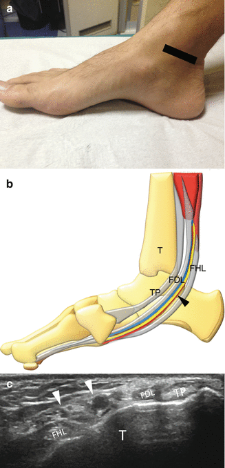
The tibial neurovascular bundle can be easily seen between the posterior tibial tendon and the flexor digitorum longus tendon (Fig. 11.2).


Fig. 11.2


Evaluation of the medial ankle to assess the tarsal tunnel. (a) The probe and patient are positioned to evaluate the tarsal tunnel on a short-axis scan. (b) Anatomical scheme of the tarsal tunnel as seen along their short axis, TP tibialis posterior tendon, FDL flexor digitorum longus tendon, FHL flexor hallucis longus tendon, arrowheads tibial neurovascular bundle, T tibia. (c) US long-axis scan of tarsal tunnel
Stay updated, free articles. Join our Telegram channel

Full access? Get Clinical Tree


