Mesothelioma is a rare cancer that is often linked to asbestos exposure. One of the biggest challenges in treating this disease has always been diagnosing it early. Because symptoms can look similar to other lung and respiratory conditions, doctors rely heavily on imaging technology to detect signs of mesothelioma and guide treatment decisions.
Over the years, imaging techniques have improved dramatically. What once required invasive procedures can now often be identified through advanced scans that provide detailed views of the body. These advancements not only help doctors diagnose mesothelioma more accurately but also help patients begin treatment sooner.
For many patients and families, a diagnosis also raises questions about past asbestos exposure and potential legal options. Some individuals choose to speak with a mesothelioma attorney co to better understand whether they may qualify for compensation related to asbestos-related illnesses while focusing on their medical care.
Why Imaging Is Important in Mesothelioma Diagnosis
Mesothelioma develops in the lining surrounding organs such as the lungs, abdomen, heart, or testes. The disease often grows slowly at first and may not cause noticeable symptoms until it has progressed.
Imaging plays a critical role by helping doctors:
- Detect abnormalities
- Identify fluid buildup
- Locate tumors
- Determine cancer stage
- Monitor treatment progress
- Guide biopsy procedures
Without imaging technology, diagnosing mesothelioma would be significantly more difficult.
The Early Days of Mesothelioma Imaging
Decades ago, doctors had limited tools available to evaluate patients with suspected mesothelioma.
Traditional Chest X-Rays
Chest X-rays were among the first imaging methods used to identify possible signs of asbestos-related diseases.
These images could reveal:
- Pleural thickening
- Fluid around the lungs
- Lung abnormalities
- Large masses
While X-rays remain useful today, they have limitations. Small tumors and early-stage mesothelioma can easily go unnoticed on a standard X-ray.
The Challenges of Early Detection
One major issue with older imaging methods was the lack of detail. Doctors often needed additional tests or invasive procedures before making a diagnosis.
As a result, many mesothelioma cases were not detected until the disease had reached an advanced stage.
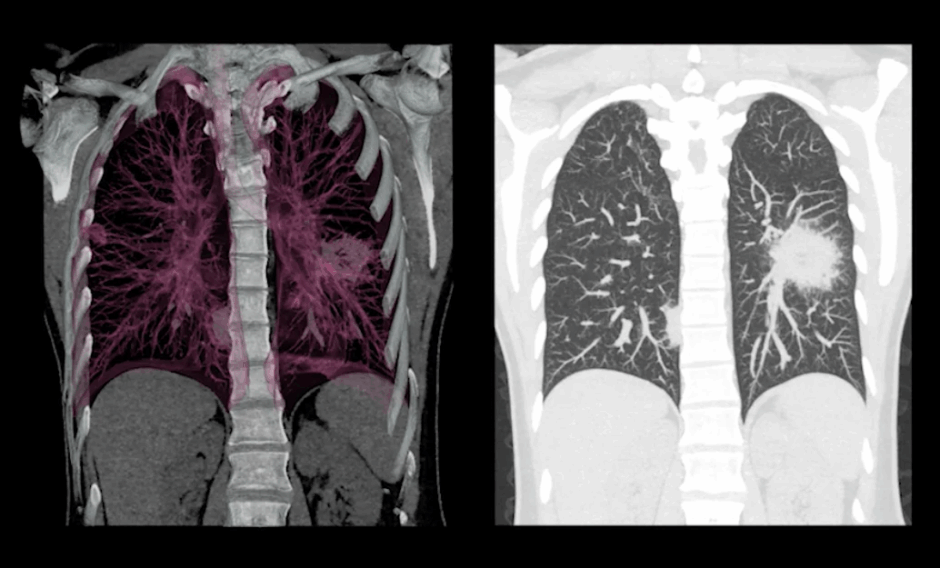
The Rise of CT Scans
The introduction of computed tomography (CT) scans marked a major advancement in cancer imaging.
How CT Scans Improved Diagnosis
Unlike traditional X-rays, CT scans create cross-sectional images of the body. This allows doctors to view structures from multiple angles and identify abnormalities more clearly.
CT scans can help detect:
- Small tumors
- Pleural thickening
- Lymph node involvement
- Fluid accumulation
- Tumor spread
Today, CT scans remain one of the most commonly used imaging tools for suspected mesothelioma cases.
Better Surgical Planning
CT imaging also helps surgeons evaluate whether a tumor can be removed and determine the safest surgical approach.
This information is essential when developing an individualized treatment plan.
MRI Technology Brings Greater Detail
Magnetic resonance imaging (MRI) introduced another level of precision to mesothelioma diagnosis.
Unlike CT scans, MRI uses magnetic fields and radio waves rather than radiation.
Advantages of MRI
MRI is especially useful for evaluating:
- Soft tissue involvement
- Chest wall invasion
- Diaphragm involvement
- Tumor boundaries
These detailed images help physicians better understand how far the disease has progressed.
When MRI Is Used
Doctors may recommend MRI when they need additional information beyond what CT scans can provide.
It is often used during staging and treatment planning rather than as the first imaging test.
PET Scans Change Cancer Detection
One of the most important developments in cancer imaging has been the use of positron emission tomography (PET) scans.
PET scans work differently from CT and MRI because they focus on metabolic activity rather than anatomy.
How PET Scans Work
Patients receive a small amount of radioactive glucose before the scan.
Cancer cells typically use more energy than normal cells, causing them to appear brighter on the images.
PET scans help doctors:
- Identify active cancer cells
- Distinguish cancer from scar tissue
- Detect metastasis
- Evaluate treatment response
Combining PET and CT Technology
Modern PET/CT scanners combine two imaging methods into one test.
This allows doctors to see both the location and activity of suspicious areas, improving diagnostic accuracy.
Advanced Imaging and Early Detection
Early diagnosis remains one of the most important factors in improving outcomes for mesothelioma patients.
New Imaging Capabilities
Modern imaging systems now offer:
- Higher-resolution images
- Faster scan times
- Improved 3D visualization
- Better tumor mapping
- Enhanced staging accuracy
These improvements allow doctors to identify abnormalities that may have been missed in the past.
Supporting Personalized Treatment
Advanced imaging helps physicians tailor treatment plans based on each patient’s specific condition.
By understanding tumor size, location, and spread, doctors can recommend the most effective therapies.
The Role of Artificial Intelligence in Imaging
While imaging equipment has improved significantly, software is also becoming more advanced.
Artificial intelligence is beginning to assist radiologists by helping identify patterns that may indicate cancer.
Potential benefits include:
- Faster image analysis
- Improved consistency
- Earlier detection of subtle abnormalities
- Enhanced treatment monitoring
Although AI is still evolving in clinical practice, it may become an increasingly valuable tool in mesothelioma diagnosis.
Imaging Beyond Diagnosis
Imaging is not only important for identifying mesothelioma. It also remains valuable throughout treatment.
Monitoring Treatment Progress
Doctors use imaging scans to determine whether:
- Tumors are shrinking
- Treatment is working
- Cancer has spread
- Additional therapies are needed
Regular imaging allows healthcare teams to adjust treatment strategies when necessary.
Long-Term Follow-Up
Even after treatment, imaging helps monitor patients for recurrence and track overall health.
This ongoing surveillance can help detect changes early and improve long-term management.
What Patients Should Expect During Imaging Tests
Many patients feel nervous before undergoing imaging procedures. Understanding what to expect can ease concerns.
CT Scan
- Usually completed within minutes
- May involve contrast dye
- Non-invasive and painless
MRI
- Longer scan time
- Requires lying still
- May feel noisy due to machine operation
PET Scan
- Includes a waiting period after tracer injection
- Generally takes longer than CT scans
- Helps identify active cancer cells
Your medical team will explain which imaging tests are most appropriate for your situation.
Conclusion
The evolution of imaging technology has transformed mesothelioma diagnosis. From simple chest X-rays to advanced PET/CT and MRI scans, doctors now have powerful tools that provide more accurate information than ever before.
These advancements help identify cancer earlier, improve treatment planning, and support better patient care throughout the disease journey. While mesothelioma remains a challenging diagnosis, modern imaging continues to play a critical role in helping physicians make informed decisions and deliver more personalized treatment strategies.
Frequently Asked Questions
What imaging test is most commonly used to diagnose mesothelioma?
CT scans are often the first advanced imaging test used because they provide detailed images of the chest and can identify abnormalities associated with mesothelioma.
Can an X-ray diagnose mesothelioma?
An X-ray may show signs of mesothelioma, but additional imaging and a biopsy are usually needed for a confirmed diagnosis.
What is the difference between a CT scan and a PET scan?
CT scans show physical structures inside the body, while PET scans highlight areas of increased metabolic activity that may indicate cancer.
Is MRI better than CT for mesothelioma?
MRI provides better soft tissue detail and may help determine how far a tumor has spread, but CT scans are typically used first.
How often are imaging scans performed during treatment?
The frequency depends on the patient’s treatment plan, but doctors commonly use imaging throughout treatment to monitor progress and evaluate results.
Related posts:
 Other Imaging Techniques in Dilated Cardiomyopathy
 Basic Echocardiography in Arrhythmogenic Right Ventricular Cardiomyopathy
 Lu-177 PSMA Therapy Cost in Germany: What Affects the Price and How to Plan Treatment
How Do Medical Reports Strengthen Personal Injury Claims?
Raising the Standard: How Nurses Can Deliver Better Hospital Care
The Health Impact of Doomscrolling: What You Should Know
Lu-177 PSMA Therapy Cost in Germany: What Affects the Price and How to Plan Treatment
How Do Medical Reports Strengthen Personal Injury Claims?
Raising the Standard: How Nurses Can Deliver Better Hospital Care
The Health Impact of Doomscrolling: What You Should Know
Stay updated, free articles. Join our Telegram channel

Full access? Get Clinical Tree


