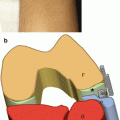
Fig. 17.1
Evaluation of the plantar hindfoot to assess the plantar fascia. (a) The probe and patient are positioned to evaluate the plantar fascia on a long-axis scan. (b) Anatomical scheme of the plantar fascia as seen along its long axis, C calcaneus. (c) US long-axis scan of the medial branch of the plantar fascia, F fat pad, arrowheads medial branch of the plantar fascia, circles enthesis of the plantar fascia, partially affected by anisotropy artifacts
Forefoot, Plantar Side
The probe must be oriented on an axial plane over the metatarsal heads. At this level, the intermetatarsal spaces and flexor digitorum tendons can be seen.
Stay updated, free articles. Join our Telegram channel

Full access? Get Clinical Tree


