Fig. 2.1
Evaluation of the anterior superior iliac spine to assess the tensor fasciae latae and the sartorius tendons and muscles. (a) The probe and patient are positioned to evaluate the anterior superior iliac spine on a short-axis scan. (b) Anatomical scheme of the anterior superior iliac spine and the tensor fasciae latae and the sartorius tendons and muscles as seen along their short axis. TFL tensor fasciae latae, ASIS anterior superior iliac spine, SA sartorius. (c) US short-axis scan of the anterior superior iliac spine, the tensor fasciae latae and the sartorius tendons and muscles
Rectus Femoris and Iliopsoas Muscles
Anatomy
The rectus femoris muscle is part of the deep group of the anterior compartment muscles of the hip, and it belongs to the quadriceps femoris complex. It inserts over the anterior inferior iliac spine (AIIS) with three different proximal tendons: the direct tendon, inserting directly on the AIIS; the indirect tendon, which is an aponeurosis into the muscle belly, running under the direct tendon proximally and then coursing externally; and the reflected tendon that anchors the chondrolabral complex of the hip joint. The rectus femoris muscle crosses the anterior aspect of the hip, coursing the anterior thigh to insert into the upper pole of the patella through the quadriceps tendon.
The iliopsoas muscle is composed of two muscles arising from the posterior abdominal wall: the psoas major and the iliacus. The psoas muscle arises from the transverse processes and the body of T12–L5 vertebrae and the lateral aspects of the discs between them; the iliacus muscle originates from the iliac fossa. The muscle belly runs obliquely down and outwards, it passes under the inguinal ligament and it inserts onto the apex of the lesser trochanter through a common tendon.
Between the joint capsule and the posterior surface of the iliopsoas muscle, there is the iliopsoas bursa, the largest synovial bursa of the human body, which communicates with the joint space in 15 % of cases.
Scanning Technique
Starting with the probe at ASIS level in horizontal position, the transducer is shifted caudally to reach the AIIS and to visualise the direct tendon of the rectus femoris muscle in its axial scan, lateral to the iliopsoas muscle. The direct and indirect tendons of the rectus femoris can be evaluated using axial and longitudinal scans. From this position, the transducer is shifted caudally to reach the muscle belly of the rectus femoris; then the probe can be rotated 90° clockwise to evaluate, by longitudinal scans, the myotendinous junctions of the rectus up to the insertion onto the AIIS.
To evaluate the iliopsoas, the probe is placed slightly medially to the AIIS, and a series of axial scans are used to detect the iliopsoas muscle. The muscle can be followed using both axial and longitudinal scans up to the insertion onto the lesser trochanter (Fig. 2.2).


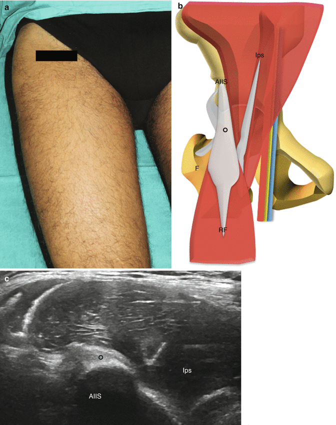
Fig. 2.2
Evaluation of the anterior thigh to assess the rectus femoris and the iliopsoas. (a) The probe and patient are positioned to evaluate the rectus femoris and the iliopsoas on a short-axis scan. (b) Anatomical scheme of the rectus femoris and the iliopsoas as seen along their short axis. AIIS anterior inferior iliac spine, circle rectus femoris conjoined tendon, RF rectus femoris muscle, Ips iliopsoas muscle, F femur. (c) US short-axis scan of the rectus femoris and the iliopsoas. The iliopsoas bursa is not visible in normal conditions
Hip Joint
Anatomy
The hip is a joint composed by the acetabulum and the femoral neck. It is a deep joint, covered by thick muscles, namely, the quadriceps and the iliopsoas. Using US, only the most superficial structures can be seen, namely, the femoral head with its articular cartilage, the anterior lateral border of acetabulum, the anterior lateral labrum and the anterior joint capsule. The intra-articular space cannot be evaluated using US.
Stay updated, free articles. Join our Telegram channel

Full access? Get Clinical Tree


