Fig. 7.1
Evaluation of the anterior knee to assess the quadriceps tendon and the suprapatellar joint recess. (a) The probe and patient are positioned to evaluate the quadriceps tendon and the suprapatellar joint recess on a long-axis scan. (b) Anatomical scheme of the quadriceps tendon and the suprapatellar joint recess as seen frontally. F femoris, P patella, arrowheads quadriceps tendon, circles suprapatellar joint recess. (c) US long-axis scan of the quadriceps tendon and the suprapatellar joint recess
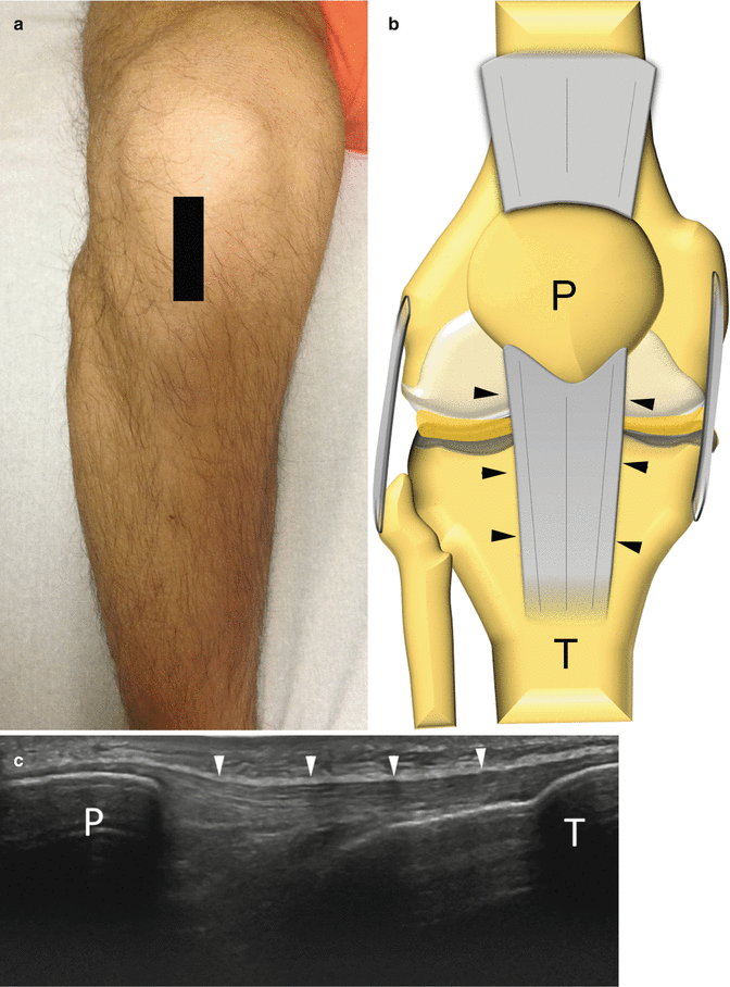
Fig. 7.2
Evaluation of the anterior knee to assess the patellar tendon. (a) The probe and patient are positioned to evaluate the patellar tendon on a long-axis scan. (b) Anatomical scheme of the patellar tendon as seen frontally. T tibia, P patella, arrowheads patellar tendon. (c) US long-axis scan of the patellar tendon
Medial Compartment
Anatomy
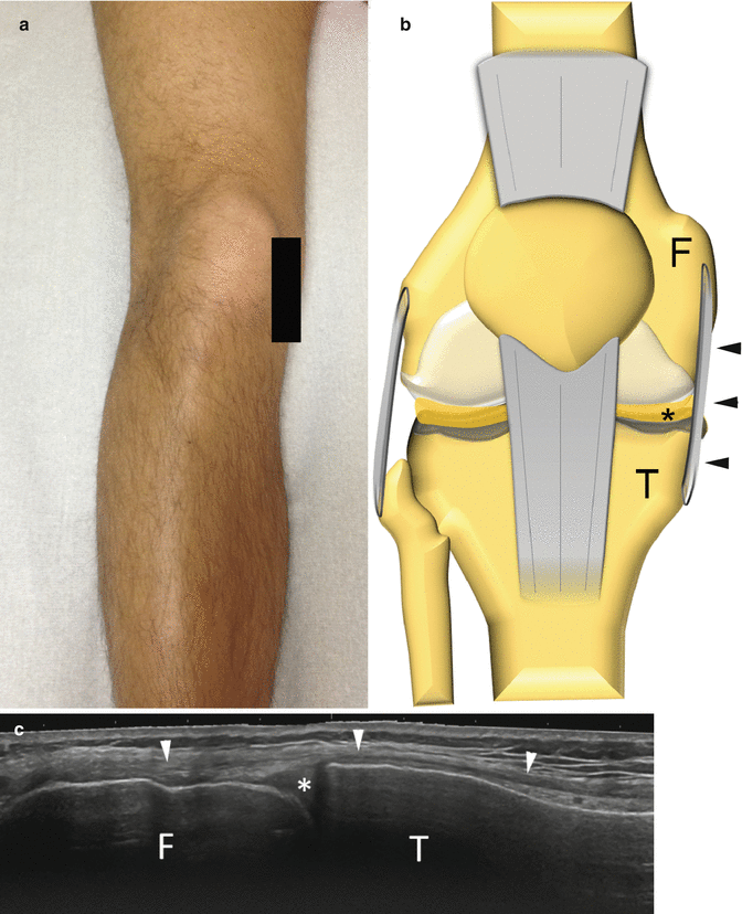
The medial collateral ligament is a band-like ligament that courses oblique from the medial aspect of the femoral condyle to the medial aspect of the tibia. Slightly anterior to the distal attachment of the medial collateral ligament, the insertion of the three tendons of the goose’s foot can be seen, namely, sartorius, gracilis, and semitendinosus. The tendons are surrounded by the goose’s foot bursa, which cannot be seen when not distended.
Scanning Technique
The knee is positioned similarly to what was reported for the examination of the anterior compartment. The probe is placed on a coronal oblique plane to detect the double-layered appearance of the medial collateral ligament. Then, the probe should be sled more distally and anteriorly to perform a longitudinal scan of the goose’s foot tendons. Of note, if the goose’s foot bursa is not distended, the tendons can be scarcely differentiated one from the other (Fig. 7.3).

