11 The musculoskeletal system
Introduction
Ultrasound is now well established in the diagnosis and initial assessment of pediatric musculoskeletal disorders. The appearance of the infant skeleton and pathology encountered in children is different to that in the adult population but the same broad principles apply. It is particularly well suited to the pediatric population as an exquisite view of the pediatric joints and cartilaginous bone can be obtained. Its prime importance in children is in the examination of the hip, both for the irritable hip and for developmental dysplasia of the hip. With recent interest in musculoskeletal sonography, a whole new field of screening for the juvenile idiopathic arthrides is developing. It is also particularly well suited to examining the soft tissues, lumps, bumps and masses that occur as a preliminary test before further more expensive imaging is undertaking.
TECHNIQUE OF SCANNING MUSCLES AND SOFT TISSUES
The highest-frequency 15L8 linear transducers are generally the best transducers to use for superficial musculoskeletal ultrasound in children. Older children may require a lower-frequency linear transducer 8L5 to visualize deeper structures and joints. All soft tissue examinations need Doppler as soft tissue hemangiomas and vascular malformations are common in children. Low flow settings on the equipment should be standard and applied, as sometimes the blood flow in these lesions is slow and difficult to detect. Power Doppler ultrasound has provided a new technique and has demonstrated hyperemia around effusions due to inflammation compared to effusions without inflammation.
Extended field of view capability is very useful when trying to demonstrate large soft tissue masses and their anatomical relationship to other structures, and this is best performed using a linear transducer with immediate view on the monitor. Any delay for reconstruction of the image is of no value in pediatric work as it prolongs the examination, often in an uncooperative and restless child. With good modern high-frequency transducers a gel stand-off is rarely required. It is extremely useful to image the contralateral unaffected limb in order to compare with the abnormal side.
The measuring package for hip angles in developmental dysplasia of the hip (DDH) is a help.
DEVELOPMENTAL DYSPLASIA OF THE HIP
The term developmental dysplasia of the hip, previously known as congenital dislocation of the hip, refers to a broad spectrum of disorders of hip development. It is now the preferred term to describe the abnormal relationship of the femoral head to the acetabulum, and it includes the varied appearance of the inadequately formed acetabulum together with complete dislocation of the femoral head as well as partial dislocation or subluxation.
The reliance on physical examination and newborn screening programs for the diagnosis has been disappointing, as dislocated hips are still diagnosed in later infancy and childhood. Late diagnosis of dislocation refers to the diagnosis being made after 3 months of age and implies a failure of neonatal detection.
Since the early description in the late 1970s of the use of ultrasound in the diagnosis of hip abnormalities, ultrasound has increasingly been used as a tool to try and improve patient outcome and to improve diagnosis of this late presentation. The ability to see the cartilaginous acetabulum and femoral head on ultrasound has made it an exceedingly attractive choice of modality for examining the infant hip. Radiography at this early age is not useful for this condition, as the femoral head and a large component of the acetabular roof has not ossified. The unossified head and labrum can be clearly seen on sonography and, in addition, a dynamic assessment can be made where the head is manipulated in the acetabulum while the sonographer watches. Also the hip can be examined in a number of scan planes, which aids in the diagnosis. Arthrography was previously used to demonstrate abnormalities such as an inverted labrum and is occasionally still used.1–3
The results of ultrasound screening have consistently shown that the non-surgical treatment rate has increased. In a Norwegian study this treatment rate was almost double in the ultrasound-screened group (34 versus 18 per 1000 infants screened) and it is assumed that many children in the ultrasound group who subsequently received treatment actually had false-positive ultrasound tests. Some would argue that this is a bad thing, as there is a risk of developing vascular necrosis of the femoral head from treatment alone.
On the other hand the surgical treatment rate did not decrease significantly in newborns screened with ultrasonography compared with those screened by physical examination alone.4
Clinical tests
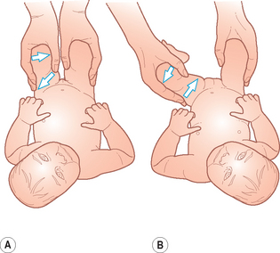
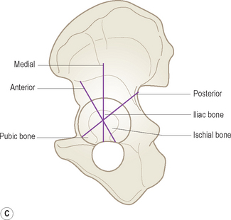
There are two clinical tests which help to identify unstable hips in newborn infants (Fig. 11.1):
• The Barlow test determines if a hip can be dislocated. The hip is flexed and the thigh adducted. With a gentle posterior pressure the femoral head can be made to move out of the acetabulum. This test actually dislocates the hip. So that when the hip is normally enlocated this test demonstrates whether the hip is partly or completely displaced out of the joint.
• The Ortolani test is the reverse of this test and relocates the head within the acetabulum. The hip is flexed and abducted and gently pulled anteriorly so that on examination a ‘click’, or the examiner feeling the dislocated femoral head moving over the acetabulum posteriorly and returning to the acetabulum, is felt. This identifies a dislocated hip and should demonstrate whether the hip is reducible.

Figure 11.1 Clinical examination for developmental dysplasia of the hip. (A) The Barlow maneuver, which is gentle adduction and a push on the knee. The hip dislocates posteriorly out of the acetabulum. (B) The Ortolani maneuver. This is the reverse so that the abducted hip is gently relocated back into the acetabulum. It typically produces the click.
These examinations should be performed by an appropriately trained health professional. There is evidence that those who specialize in hip abnormalities have a better outcome with fewer late presenters. However there are those who argue that some infants are normal at birth (and therefore screening) and then go on to develop dislocation.
There are a number of risk factors associated with developmental dysplasia of the hip:
• Family history of congenital dislocation of the hip is the most important risk factor. In some series of DDH up to 20% of infants treated had a family history.
• Female firstborn and pregnancies with oligohydramnios.
• A foot deformity that requires further treatment.
• A clicky hip as discovered on the Barlow or Ortolani test.
All those infants with displaced or unstable hips identified at screening and with a positive screening test should be seen by an orthopedic surgeon experienced in the field. It is very important to identify unstable hips as soon as possible, as the tissues are still supple and the chance of successful treatment is better at an earlier age.
Ideally the orthopedic surgeon will perform a joint clinic with the sonographer or have access to a hip ultrasound service. This combined approach is undoubtedly the most successful and rewarding for the sonographer and will ultimately yield the best results. Also in this milieu of increasing litigation the security of operating as a team is beneficial to all, including the patient.
Infants with displaced or unstable hips should be examined by 2 weeks of age and for those at risk the examination is best performed when an infant is 4–6 weeks old. By this time, most immature hips have stabilized.
It is very important that the parents are kept informed and made aware that any screening program may not identify all affected children.
There are other classical signs of congenital hip displacement which may be present at birth and which become increasingly apparent after the first 6 weeks as the legs extend and the head of the femur displaces upwards. Parents should be made aware of
• Limb shortening. This is most apparently when the hips are flexed and the levels of the knees are compared.
• Leg posture. The thigh on the affected side tends to be held in partial lateral rotation, flexion and abduction.
• Thigh asymmetry. Different skin creases on the inside of the thigh may be apparent on the affected side.
• Limited abduction. In a supine position with the hips flexed, abduction may be limited as the femoral head remains dislocated.
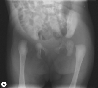
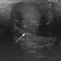
Once the child is walking, other suspicious features are a limp or anxieties about the child’s walk, discrepancy in leg length and abnormalities of lower limb posture (Fig. 11.2).

Figure 11.2 Late development of developmental dysplasia of the hip (DDH). This radiograph demonstrates the appearances of the late presentation of a dislocated hip. The acetabulum is steep and shallow and the femoral head is dislocated superiorly and posteriorly. The femoral capital epiphysis is also smaller than that on the left. The dislocated hip demonstrates how these children may present with apparent shortening of the leg on the affected side.
TECHNIQUE OF HIP SCANNING
There is no universally accepted standard technique for ultrasound examination of the infant hip in the world today, and there are at least three described methods practiced and in use:
• Graf has devised a complex classification based on the acetabular structure and position of the femoral head. Several lines and angles are measured and a single coronal image is produced. There is no dynamic (i.e. scanning and watching the femoral head movement while under stress) component to his technique. A simplified version of his technique is probably the most widely practiced method in Europe: for example, Rosendahl and colleagues have adapted Graf’s method so that it assesses hip morphology and hip stability separately, to distinguish their relative importance for treatment outcome.
• Dynamic sonography of the infant hip, as described by Harcke, is a dynamic examination which stresses positional relationships and stability.
• The third technique is simply a line drawn down from the baseline which assesses what percentage of the femoral head is lying within the acetabulum.
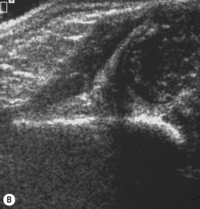
Undoubtedly the best results are obtained with the sonographer working in close collaboration and conjunction with an orthopedic surgeon experienced in DDH so that consistent reliable results are produced and terminology understood by all, with improved outcomes for the infant.
The infant must be relaxed for the examination so that adequate assessment of the hip can be made and so that stress maneuvers are accurate. A screaming kicking infant cannot be examined and it is essential that they are given time to feed and calm down. A soother dipped in 5% dextrose water is invaluable.
Curvilinear probes can be used, but it is now widely accepted that high-frequency linear probes are undoubtedly the best and do not distort the image for angle measurements if these are to be taken. For a young infant a 15L8 can be used and a lower frequency will be needed in an older infant. Once the femoral head starts ossifying and produces a densely acoustic shadow, sonography is less reliable, but this is generally later and after 3–6 months. When the ossific nucleus enlarges, the acetabular floor is obscured by its acoustic shadow, but by this time plain radiographs are reliable.
The Graf technique
The Graf technique requires the sonographer to be able to identify important landmarks in the coronal plane and from these landmarks measure the alpha and beta angles. This technique requires some training and practice and the sonographer should ensure that they are adequately trained by an experienced practitioner in this method if it is to be used.5
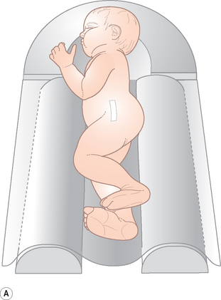
It is recommended that a positioning device is used to immobilize the patient. The infants are placed on their side and the transducer is placed over the greater trochanter (Fig. 11.3). The hip should be in slight flexion. When the trochanter, femoral neck and acetabulum lie in the same plane, this is optimal. The transducer is positioned so that it lies exactly in the frontal plane parallel to the body’s long axis and should not be tilted. The standard sonographic plane should be easily achieved in this position.


Figure 11.3 The Graf cradle. (A) The infant is positioned on its side with hip in slight flexion for the Graf method of examining the hip. (B) Images of the baseline either too anterior or posterior (from left to right). (C) Schematic diagram of the incorrect plane of the image.
The following main reference points need to be identified:
• the cartilaginous portion of the acetabular roof
• the bony promontory of the superior bony acetabular rim
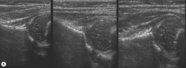
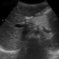
Once this standard plane has been achieved, the reference lines are drawn (Figs. 11.4 and 11.9).

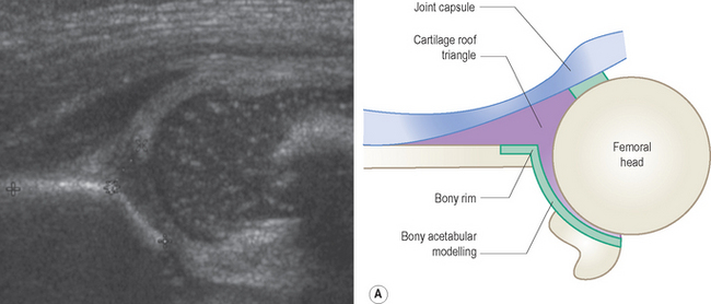
Figure 11.4 Graf’s—standard plane. From this, the standard reference lines are drawn and the hip angles obtained. See also Fig. 11.9
The alpha angle is described as the bony roof angle and is the most important to measure. The beta angle is the cartilaginous roof angle and is not universally used (Table 11.1).
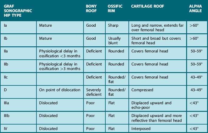
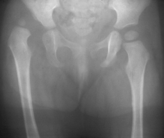
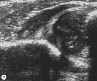
Table 11.1 is an adaptation of Graf’s classification of hip types. Types III (subluxation) and Type IV (dislocation) are probably the easiest to identify. Graf Type III involves persistent lateral displacement of the femoral head from the acetabular floor with deviation of the labrum. Type IV hips are complete dislocations, usually posterior and towards the head, with the femoral metaphysis obscuring the acetabular floor (Fig. 11.5).


Figure 11.5 Dislocated hip. (A) This plain radiograph demonstrates the abnormalities to be detected on ultrasound. The right hip is completely dislocated. The acetabulum is steep and short and there is insufficient cup to keep the femoral head within the socket. On the left the acetabulum is normally short and steep although the head is not as severely dislocated as on the right side. (B) Ultrasound examination of the right hip shows how the head is completely dislocated posteriorly. The normal coronal plane cannot be produced. (C) Coronal sonogram of the left hip. The left hip is decentered, also lying predominantly out of the acetabulum.
From a sonographer’s point of view the standard image plane cannot be achieved when the head is dislocated, neither can accurate angles be measured. Types III and IV are very rarely detected outside specialist centers.
• Type I are the mature hips with a deep acetabulum and angles over 60°.
• Type II a are those under 3 months of age with a shallow acetabulum and alpha angles of 50–59°. These are considered to be physiologically immature hips but stable.
• Type II b are the infants of over 3 months who have shallow acetabulae with alpha angles of between 50 and 59° who are also considered by Graf to be inherently stable.
• The most important group to identify for the sonographer are the unstable Type II c with shallow acetabulae and angles of 43–49°. These are considered to be unstable and require immediate treatment.
The Graf classification is complex, and training is needed in order to be competent in the technique.

Stay updated, free articles. Join our Telegram channel

Full access? Get Clinical Tree

