(1)
Princess Elizabeth Hospital Le Vauquiedor St. Martin’s Guernsey, Channel Islands, UK
(2)
Brighton and Sussex Medical School, Brighton, England UK
Abstract
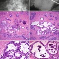
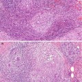
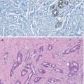
The breast tissue develops from the milk line from approximately 7 weeks gestation. The ducts arise from the ectodermal mammary ridge which develops into a budding stage by the 12th week. The milk line extends from the axilla to the groin and is responsible for vestigial accessory nipples. However the axilla to groin concept of the nipple line in humans has been challenged as accessory nipples are only found in the axillo-pectoral region (Mansel et al. 2009). The most common embryological vestige is accessory breast in the axilla (Fig. 1.1).
Learning Points
The breast tissue develops from the milk line.
At birth there is no difference between male and female breasts.
Pubertal growth is due to glandular and fibrofatty proliferation.
The amount of fibroglandular tissue decreases with age.
Benign breast disease and cancer arise in the terminal duct–lobular unit.
Multiple hormonal stimulation significantly increases the volume of the breast tissue during pregnancy.
1.1 Embryology of the Breast
The breast tissue develops from the milk line from approximately 7 weeks gestation. The ducts arise from the ectodermal mammary ridge which develops into a budding stage by the 12th week. The milk line extends from the axilla to the groin and is responsible for vestigial accessory nipples. However the axilla to groin concept of the nipple line in humans has been challenged as accessory nipples are only found in the axillo-pectoral region (Mansel et al. 2009). The most common embryological vestige is accessory breast in the axilla (Fig. 1.1).
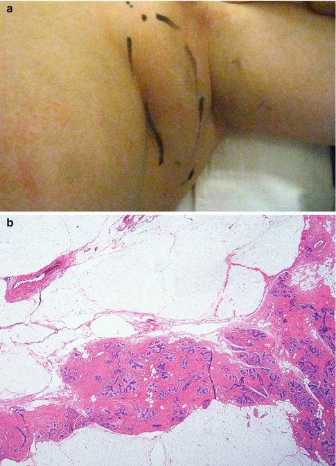
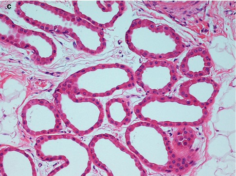
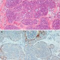
Fig. 1.1
(a) Accessory breast tissue which presented as an axillary mass. (b) The histology shows normal breast lobular units. (c) The presence of apocrine sweat glands in the surrounding tissue confirmed axillary origin of the breast tissue
Whist developing in utero, the breast epithelium and stroma are under the influence of growth factors and placental hormones, which include prolactin, oestrogen and progesterone (Forsyth 1991). Falling of maternal oestrogens in the neonatal period stimulates the production of prolactin which induces secretory changes in the breast. This results in unilateral or bilateral enlargement of the breasts which may be associated with secretion of ‘witches milk’. At birth there is no difference between the male and female breast tissue, but the presence of testosterone leads to the involution of the male breast.
1.2 Pubertal Changes
Thelarche, which commences between 10 and 12 years, heralds the rapid growth of the breast at the onset of puberty. The pituitary hormones follicle-stimulating hormone (FSH) and luteinising hormone (LH) stimulate the production of oestrogen and progesterone, resulting in the enlargement of the areola and the nipple, development of the lobules and associated acini and increase in intralobular stroma and fat. Abnormal secretion of the hormones can result in premature thelarche which is characterised by unilateral or bilateral development of breast tissue before puberty. Erroneous interpretation of breast enlargement in premature thelarche as pathological can lead to surgical excision rendering the child amastic. The breast tissue in premature thelarche regresses with time.
1.3 Gross Anatomy of the Adult Female Breast
Related posts:

Stay updated, free articles. Join our Telegram channel

Full access? Get Clinical Tree


