Radiologists are often required to evaluate postoperative spine imaging to assist the surgeon with further clinical management. This article reviews common spine surgical techniques and their proper evaluation on imaging from a surgical perspective. The article attempts to provide a basic surgical foundation for radiologists and a clearer delineation of important points and complications that should be commented on when evaluating the postoperative spine on imaging.
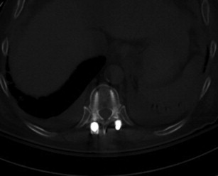
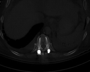
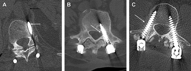
When evaluating posterior spinal hardware special attention should be given to pedicle screws, which are often used as anchoring devices in spinal fusion and stabilization procedures. Optimal pedicle screw placement is when the screw traverses the central portion of the pedicle and is parallel to the vertebral endplate ( Fig. 6 ). Complications include medial angulation of the screw, which may lead to disruption of the medial cortex of the pedicle and nerve root irritation. Lateral angulation may also occur and is especially important in the cervical spine, where the screw may traverse the foramen transversarium and potentially compromise the vertebral artery. Improper placement may also lead to screws traversing the anterior cortex of the vertebral body, sometimes abutting the aorta. Similarly, when evaluating anterior plate and screw fixation the screws may overpenetrate and lead to dural, cord, or nerve root injury ( Fig. 7 ). Such complications can be avoided by using intraoperative fluoroscopy and pedicle screw electrical stimulation to confirm appropriate screw positioning.
When evaluating posterior spinal hardware special attention should be given to pedicle screws, which are often used as anchoring devices in spinal fusion and stabilization procedures. Optimal pedicle screw placement is when the screw traverses the central portion of the pedicle and is parallel to the vertebral endplate ( Fig. 6 ). Complications include medial angulation of the screw, which may lead to disruption of the medial cortex of the pedicle and nerve root irritation. Lateral angulation may also occur and is especially important in the cervical spine, where the screw may traverse the foramen transversarium and potentially compromise the vertebral artery. Improper placement may also lead to screws traversing the anterior cortex of the vertebral body, sometimes abutting the aorta. Similarly, when evaluating anterior plate and screw fixation the screws may overpenetrate and lead to dural, cord, or nerve root injury ( Fig. 7 ). Such complications can be avoided by using intraoperative fluoroscopy and pedicle screw electrical stimulation to confirm appropriate screw positioning.

