3.2 2D Ultrasound
3.2.1 Transabdominal Ultrasound
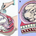
The first stage of labor presents a series of technical challenges for transabdominal 2D sonographic imaging (Fig. 3.2). At this stage, the ossified fetal skull lies deep in the bony pelvis and shadows the posteriorly located uterine cervix; In addition, during labor the cervix effaces and dilates further impairing its sonographic imaging. Due to these limitations, the use of transabdominal ultrasound in the first stage of labor has been restricted to the imaging of the fetal presenting part, its intracranial structures, and their relation to maternal to assess fetal head pelvic engagement (Fig. 3.3a–c) and fetal head position.
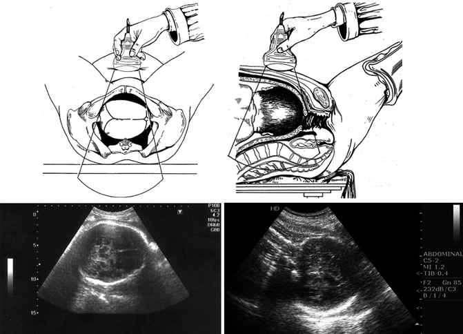
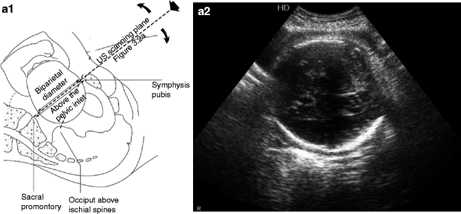
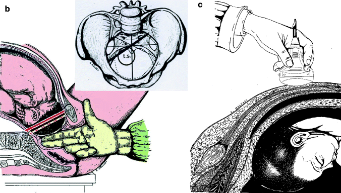
Fig. 3.2
Axial (a) and longitudinal (b) transabdominal intrapartum ultrasound
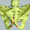
Fig. 3.3
(a) Diagram depicting the ultrasonographic assessment of head engagement. The dotted line depicts the scanning plane at the level of the inlet used to determine the engagement of fetal head. A fetal biparietal diameter at or below pelvic inlet sign fetal head engagement (Sherer et al. 2003 [5]). (b) Sign of Farabeuf: normal engagement of the fetal head in synclitism, in which it is observed that the biparietal diameter (dashed line) is parallel to the conjugated diagonal (red line). With its narrow upper diameters: the thick line represents the transverse diameter left (t.s.), on which engages more frequently the fetal head (dotted line). (c) The image demonstrates the commitment of the fetal head during the first stage of labor in synclitism by transabdominal ultrasound
Sherer et al. [7] compared transabdominal ultrasound and digital examination to assess fetal head engagement in 222 patients in labor. The pelvic inlet was established as an oblique line from the symphysis pubis to L5-S1 vertebra. Fetal head engagement was determined if the biparietal diameter (BPD) was at or below the line. The ultrasonographic assessment shows agreement with digital examination in most cases (85.6%. 95% CI, 80.8–90.3). When nulliparous and parous patients were compared, the correlation between digital and sonographic assessment remains high (nulliparous 81.5%. 95% CI, 73.4–88.0, and parous 90.3%. 95% CI, 84.1–95.9). In another study [8], the same group compared ultrasonographic determination of fetal head position, defined as the relation of fetal occiput to maternal longitudinal axis, to digital examination in 102 patients in the first stage of labor. Transabdominal ultrasound was used to image midline intracranial structures (cavum septum pellucidum, falx cerebri, thalami, and cerebellar hemispheres) and cranial structure (eyes, nasal bridge, and cervical spine) to determine fetal head position. Results were compared to digital examinations classified as occiput anterior (OA) (Fig. 3.4), posterior (OP) (Fig. 3.5), left or right occiput transverse (Fig. 3.6) (LOT) (ROT), and left or right occiput anterior or posterior (LOA) (ROA) (LOP) (ROP). Ultrasonographic and digital examination correlation was poor and agreed in only 24% of patients (95% CI, 16–33) [8]. Similar results were presented by Akmal et al. [9] in a prospective study including 496 singleton pregnancies in labor at term that compared digital examinations to transabdominal ultrasound. In this study, digital examination was considered correct if the fetal head position was within 45° of the ultrasound imaging. They found that digital examination failed to define the fetal head position in 166 (33.5%) cases and in the 330 (66.5%) cases where the position was determined correctly. The findings of the digital and sonographic examinations were in full agreement in only 163 (49.4%) cases. The rate of correct identification of the fetal position by digital examination increased with cervical dilatation, from 20.5% at 3–4 cm to 44.2% at 8–10 cm. The authors concluded that digital examinations fail to identify the correct position of fetal head in the majority of cases when compared to transabdominal ultrasound (Fig. 3.7).
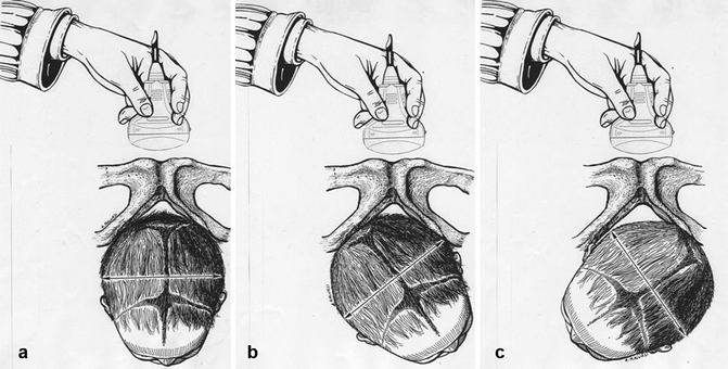
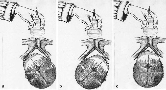
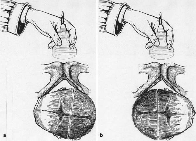
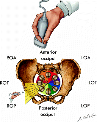
Fig. 3.4
The image demonstrates the anterior occiput position of the fetal head: (a) median anterior occiput position, (b) right anterior occiput position, and (c) left anterior occiput position
Fig. 3.5
The image demonstrates the posterior occiput position of the fetal head: (a) left posterior occiput position, (b) right posterior occiput position, and (c) median posterior occiput position
Fig. 3.6
The image demonstrates the transverse occiput position of the fetal head: (a) left transverse occiput position and (b) right transverse occiput position
Fig. 3.7
Fetal occipital position in the pelvis during labor and corresponding presentation indices. The indices of presentation are distant 45° degrees from each other. The dotted lines between LOA, LOT, and LOP indicate that between an index and the other one can determine other different degrees of position index. In case some are more prone to the right rear position, while others transverse to the right
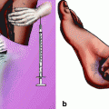
3.2.2 Translabial and Transperineal Ultrasound
Before the advent of transvaginal probes, translabial and transperineal ultrasound was used to image the uterine cervix using abdominal transducers. To obtain a transperineal picture the patient is placed in a dorsal lithotomy position with the hips flexed and slightly abducted. A standard abdominal curved-array transducer covered with a plastic wrap or a glove is positioned in the midsagittal plane against the symphysis pubis. The transperineal and translabial approach bypasses the pelvic bone structures providing unobstructed access to pelvic soft tissues imaging, the resulting image includes (anterior – posterior) the symphysis pubis, the bladder and urethra, vagina, cervix and rectum, and cranially the fetal presenting part and pelvic walls (Fig. 3.8).
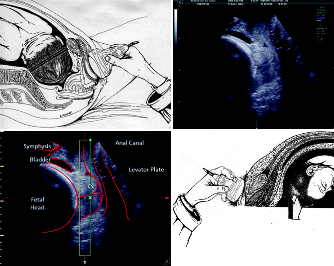
Fig. 3.8
Intrapartum translabial 2D ultrasound imaging maternal pelvis and fetal head and anatomical landmarks
Zilianti et al. [10
 General Intrapartum Sonography Setup and Use in Labor
 Intrapartum Sonography and Clinical Risk Management
 New Technologies for Monitoring Labor Progress
 Clinical Evaluation of Labor and Intrapartum Sonography
 Occiput Posterior Position and Intrapartum Sonography
 Intrapartum Sonography and Labor Progression


Related posts:
General Intrapartum Sonography Setup and Use in Labor
Intrapartum Sonography and Clinical Risk Management
New Technologies for Monitoring Labor Progress
Clinical Evaluation of Labor and Intrapartum Sonography
Occiput Posterior Position and Intrapartum Sonography
Intrapartum Sonography and Labor Progression
Stay updated, free articles. Join our Telegram channel
Full access? Get Clinical Tree
