CHAPTER 4 The white lung field
4.1 Collapse
Check the position of the oblique and horizontal fissures (pp. 13 and 14). Any displacement from their normal position suggests collapse. Collapse of any of the lobes of the lung gives a distinct appearance on the X-ray.
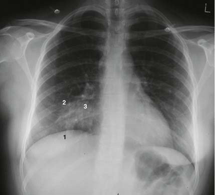
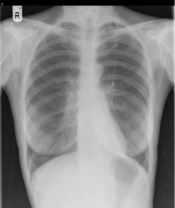
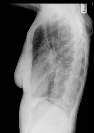
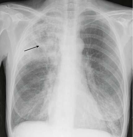
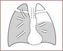
1. Right upper lobe collapse. There is an area of whiteness in the upper zone of the right lung (1). The horizontal fissure is elevated, there is an apparent right upper hilar ‘mass’, the trachea is deviated to the right (2) and the ribs over the area of whiteness are closer together than is normal. On the lateral film the increased whiteness in the uppermost part of the chest may be seen.
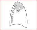
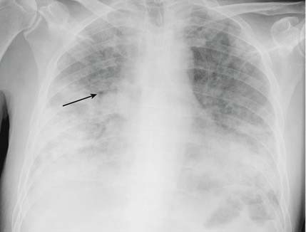
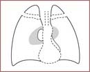
2. Right middle lobe collapse. This can be difficult to spot. The right diaphragm may be slightly raised (1) and the horizontal fissure (2) may be lower than usual. The upper part of the lower zone may have a hazy white appearance (3) and the heart border is sometimes indistinct. It is easier to detect in the lateral film. There is a triangular area of whiteness with its apex at the hilum (4) and its base running between the sternum and the diaphragm (5).
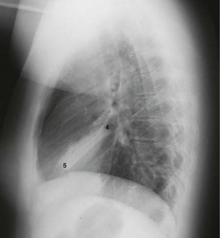
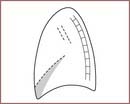
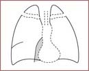
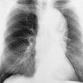
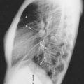
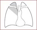
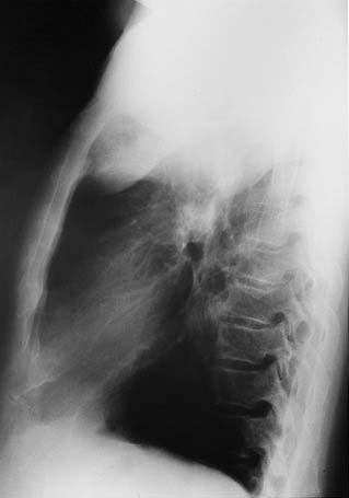
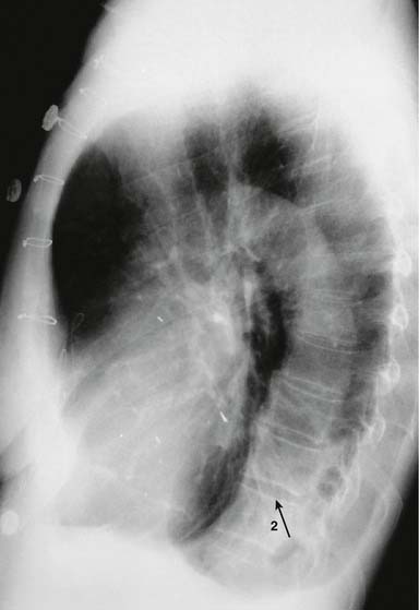
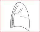
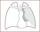
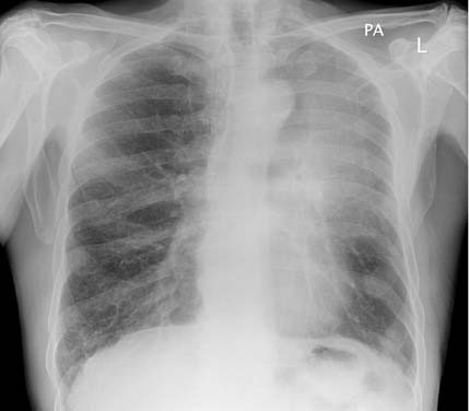
3. Right lower lobe collapse. There is a whiteness immediately above the diaphragm (1) causing a loss of its outline. On the lateral film there is a white triangle at the lower posterior part of the lung field (2). Note how the outline of the right heart border is maintained.
4. Left upper lobe collapse. This is difficult to spot. Remember that most of the left upper lobe lies in front of, as opposed to above, the left lower lobe. When it collapses it causes a haze to appear over the whole of the left lung field.
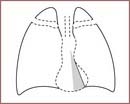
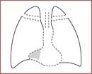
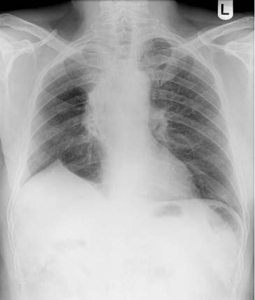
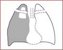
5. Left lower lobe collapse. This is easy to miss. The left lower lobe collapses down behind the heart. The left lung field appears much darker than normal and the heart shadow will appear much whiter than normal. If you look carefully you can see a white triangle behind the heart (1). On the lateral film you may see a white triangle at the bottom posterior corner of the lung fields (2) and the vertebral bodies will appear whiter.
4.2 Volume loss
A pneumonectomy is another cause of a white lung. You should know from the history and your examination that the patient has had a pneumonectomy. Look at the X-ray for the following features:
A very rare cause of a similar appearance is extensive hypoplasia or congenital absence of one lung.
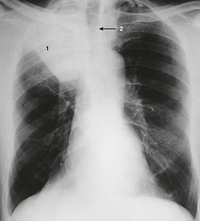
This patient had right upper lobectomy and postoperative radiotherapy. Both of these have led to volume loss in the remaining right lung. The trachea has been pulled to the right as a result. The lung is of increased blackness on the right compared to the left because the remaining lung is hyperinflated.
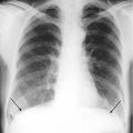
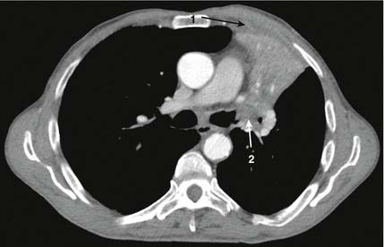
Tracheal deviation can be the result of it being pushed by a mass lesion in the mediastinum, most often an enlarged thyroid gland, as in the case shown here. The lung volumes in this case are normal, and the ribs and diaphragms are in their normal positions. In the elderly a very tortuous aorta may also lead to tracheal shift.
4.3 Consolidation
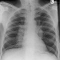
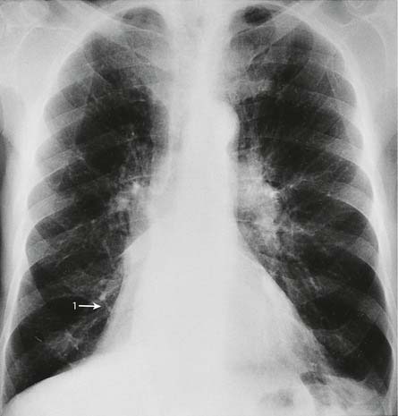
This is the appearance of a lobar pneumonia. Notice how the inferior margin of the consolidation is quite straight. This appearance indicates a right upper lobe pneumonia. An air bronchogram (arrow) is visible.
This shows much more widespread consolidation affecting both lungs, especially in the mid to lower zones, and is a bronchopneumonia. An air bronchogram (arrow) is again visible.
Again you can see an area of white lung. Look first at the nature of the whiteness and its border. If it is uniform with a well-demarcated border you are much more likely to be dealing with an area of collapse or a pleural effusion. If the shadowing is not uniform and the border is not so well demarcated the possibilities are consolidation, fibrosis or some other infiltrative condition. It can be difficult to diagnose consolidation so make your way carefully through the following steps:
If a patient is admitted with pneumonia it is not necessary to repeat the chest X-ray before discharge if they make a satisfactory recovery. For patients who fail to recover, further X-rays are necessary to look for possible complications such as the development of an empyema or pulmonary abscess. For patients ill enough to warrant admission to intensive care, early follow-up X-rays are warranted to ensure that their condition is not progressing.
4.4 Pneumocystis carinii (jiroveci) pneumonia (PCP)
Pneumocystis carinii pneumonia (PCP) can be difficult to diagnose on a chest X-ray and in 10% of patients with PCP the chest X-ray is normal. It is something to suspect if a patient presents with shortness of breath and hypoxia which are out of proportion to a relatively normal looking chest X-ray.
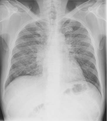
4.5 Pleural effusion
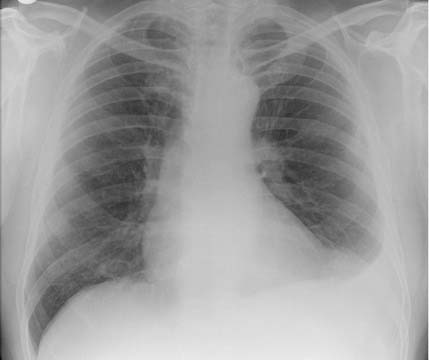
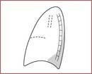
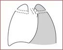
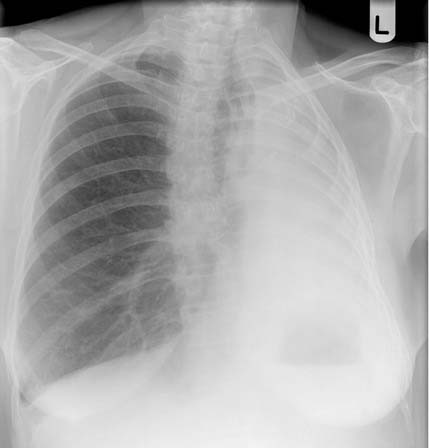
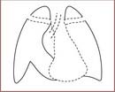
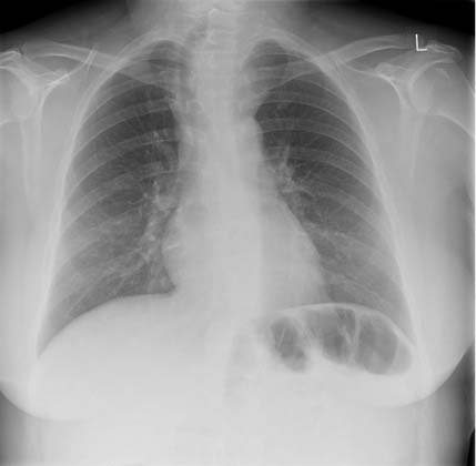
This pair of films shows the varied appearances of pleural effusions. The first film shows a small left effusion filling the costophrenic angle. It has a curved upper margin.
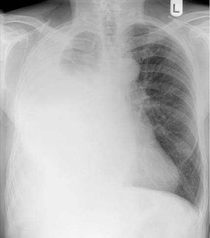
If you see an area of whiteness at the base of a lung then the possible causes are a pleural effusion, a raised hemidiaphragm and an area of consolidation or collapse. You need to determine which of these it is.