Criteria
Description
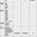
Clinical criteria
Patches and plaques plus lesions in a non-sun-exposed location, size/shape variation of lesions, and poikiloderma; 1 point for 1 factor, 2 points for 2 or more factors
Histopathologic criteria
Superficial lymphoid infiltrate present plus epidermotropism without spongiosis and lymphoid atypia (1 point for 1 factor, 2 points for 2 factors)
Molecular-biological criteria
Clonal TCR gene rearrangement is present
Immunopathologic criteria
Fewer than 50 % of the T cells express CD2, CD3, or CD5, <10 % of the T cells express CD7, and there is discordance of epidermal and dermal cells with expression of CD2, CD3, CD5, or CD7 (1 point for any of these present)
MF has its own unique staging system (Table 2) [14]. Thirty percent, 35, 20, and 15 % of patients present with T1, T2, T3, or T4 disease, respectively.
Table 2
Mycosis fungoides TNM classification and staging system for cutaneous T-cell lymphoma
T (skin) classification | |
T1 | Limited patch or plaque on <10 % of the skin surface |
T2 | Generalized patch or plaque on >10 % of the skin surface involved |
T3 | Tumorous skin involvement |
T4 | Erythroderma |
N (lymph nodes) classification | |
N0 | No clinically abnormal lymph nodes |
N1 | Clinically abnormal lymph nodes with histopathology Dutch grade 1 or NCI LN0-2 |
N2 | Histopathology Dutch grade 2 or NCI LN3 |
N3 | Histopathology Dutch grades 3–4 or NCI LN4 |
M (visceral organs) classification | |
M0 | No visceral involvement |
M1 | Visceral involvement |
B (blood) classification | |
B0 | No significant blood involvement with ≤5 % Sézary cells |
B1 | Low blood tumor burden |
B2 | High blood tumor burden with positive clone plus one of the following: ≥1,000/μL Sézary cells, CD4/CD8 ≥ 10, CD4+CD7− cells ≥40 %, or CD4+CD26− cells ≥30 % |
Clinical stage | |
IA | T1N0M0B0-1 |
IB | T2N0M0B0-1 |
IIA | T1-2N1-N2B0-1 |
IIB | T3N0-2M0B0-1 |
IIIA | T4N0-2M0B0 |
IIIB | T4N0-2M0B1 |
IVA1 | T1-4N0-2M0B2 |
IVA2 | T1-4N3M0B0-2 |
IVB | T1-4N0-3M1B0-2 |
Early-stage (IA–IIA) MF has a favorable prognosis with a median survival of 13 years [12] and is generally treated with skin-directed therapies, including topical corticosteroids, topical chemotherapeutic agents (nitrogen mustard and carmustine), topical retinoids (stage IA only), RT (local electron-beam RT for unilesional or total skin electron-beam RT for more extensive and progressive skin disease), and phototherapy using ultraviolet B (UVB) or psoralen + ultraviolet A photochemotherapy (PUVA). No specific treatment is preferred over the others; however, trying a different regimen is recommended on progression after initial treatment. In a study of 103 patients randomized to a combination chemotherapy (cyclophosphamide, doxorubicin, etoposide, and vincristine) and 30 Gy of total skin electron-beam therapy (TSEBT) vs. sequential topical therapy, there was no significant difference in disease-free or overall survival [15]. Because of the morbidity of TSEBT, it is generally reserved for progressive lesions or thicker tumor plaques.
Patients with more advanced MF (stage IIB–IV) have a worse prognosis, with an overall survival rate of 3.5–4 years for stage IIB–III and 1.5 years for stage IV and require more aggressive treatment regimens [12].
Limited stage IIB disease can be treated with a combination of local RT to the tumors with other topical agents applied to adjacent plaques or systemic therapy, while more extensive stage IIB and IIIA disease can be treated with systemic retinoids, interferon, histone deacetylase inhibitors, Denileukin diftitox, systemic chemotherapy, and TSEBT. Generally TSEBT is only used in stage IIB disease, because patients with more advanced disease may suffer severe desquamation after doses as low as 4 Gy.
Stage IIIB and IV requires systemic therapy to target malignant cells in the blood and may include extracorporeal photochemotherapy, systemic retinoids, interferon, histone deacetylase inhibitors, Denileukin diftitox, methotrexate, or allogeneic stem cell transplantation. These systemic agents can be combined with skin-directed therapies.
Table 3
Acronyms
NHL | non-hodgkin lymphoma |
PCTCL | primary cutaneous T-cell lymphoma |
PCBCL | primary cutaneous B-cell lymphoma |
LDH | lactate dehydrogenase |
CT | computed tomography |
PET |


