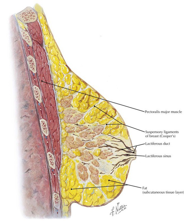
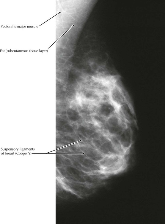
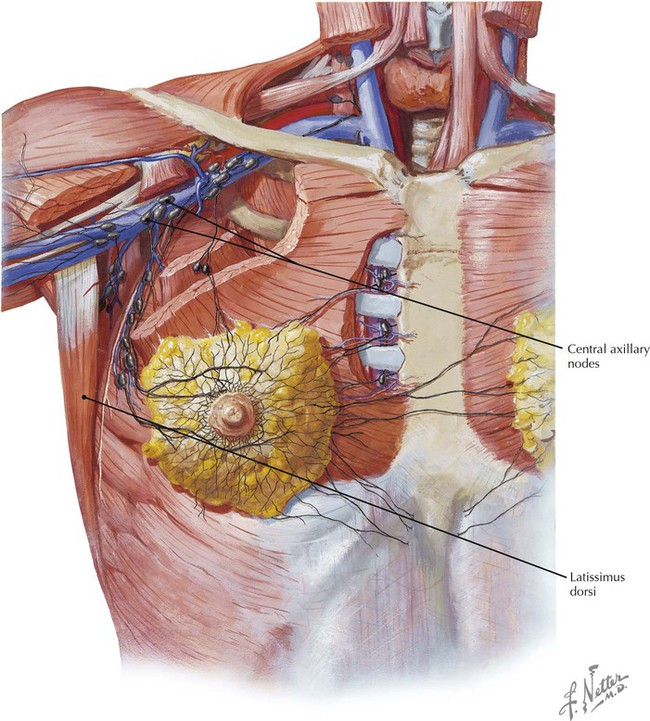
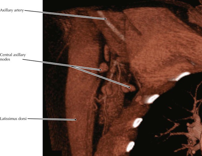
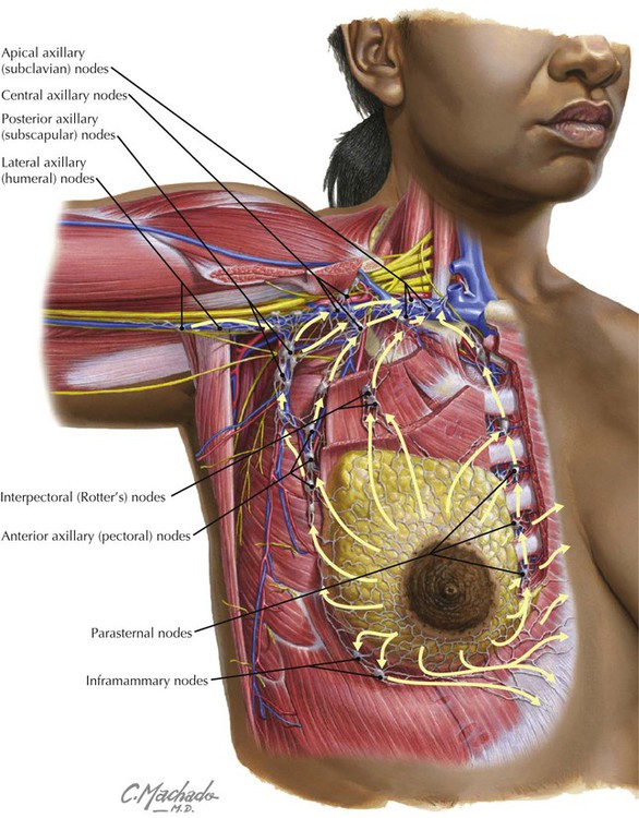
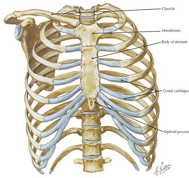
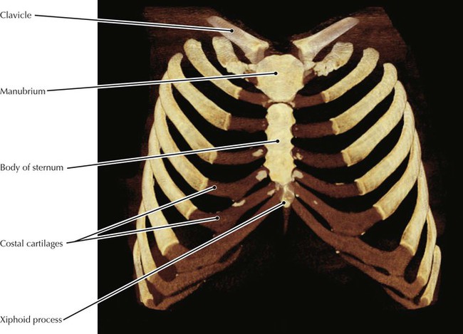
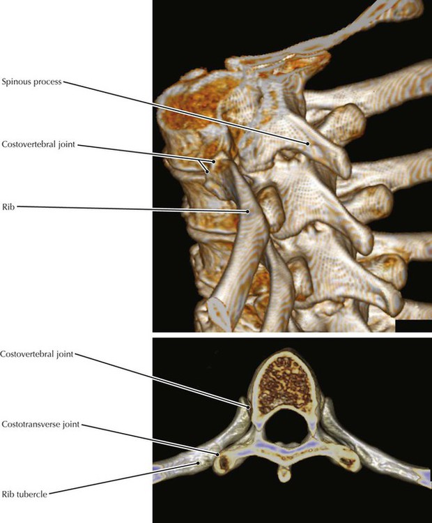
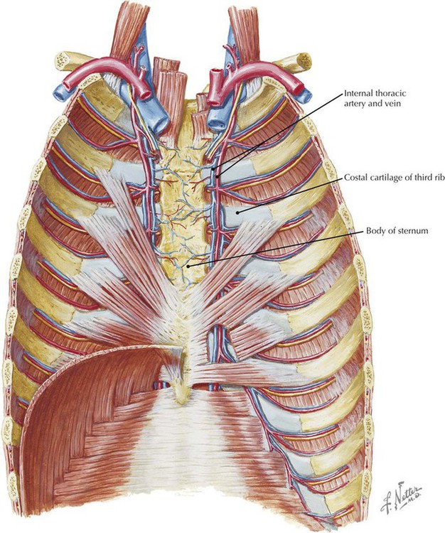
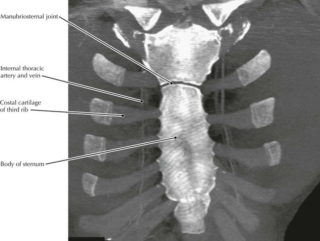
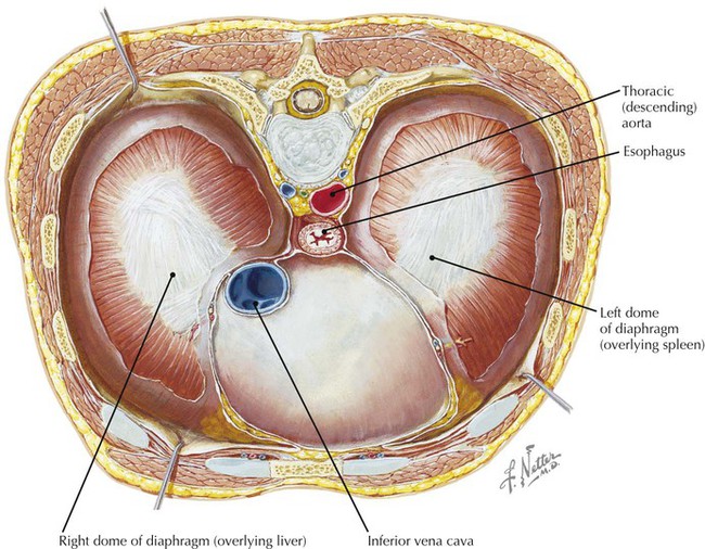
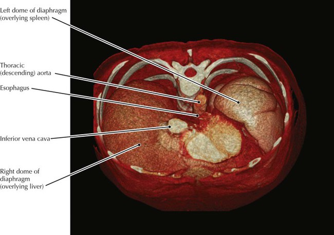
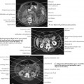
• Standard projections for screening mammography are the MLO view shown above and a craniocaudal (CC) projection. • When clinical breast examination reveals a suspicious finding, diagnostic mammography should be requested. Sometimes, routine screening MLO and CC views are not adequate for visualization of a mass, so additional mammographic views such as spot compression, magnification, and 90-degree mediolateral ones are performed and are often followed by ultrasonography. • Cooper’s ligaments appear in mammograms as very thin white lines. • The arm is elevated in this patient. • In subclavian venous puncture for central line placement the vein initially punctured is technically the axillary vein, which becomes the subclavian at the first rib. Thus, it is clinically important that the axillary vein lies anterior and inferior (i.e., superficial) to the axillary artery and the cords of the brachial plexus. • The internal thoracic (mammary) artery and vein give rise to the anterior intercostal vessels, which anastomose with the posterior intercostal vessels, which are branches of the thoracic aorta. • The joints between the costal cartilages and the ribs are classified as primary cartilaginous joints (synchondroses), whereas the joint between the manubrium and the sternum is a secondary cartilaginous joint (symphysis). • The diaphragm is innervated by the phrenic nerve, which is typically composed of segments from the ventral rami of the C3, C4, and C5 spinal nerves. • Because the supraclavicular nerves also receive innervation from C3 and C4, pain from much of the diaphragm is referred to the shoulder region. • The liver and spleen are partially protected from injury from the lower part of the rib cage as seen in this CT image.
Thorax
Breast, Lateral View
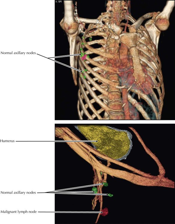
Lymph Nodes of the Axilla
Internal Thoracic Artery, Anterior Chest Wall
Diaphragm
Left Lung, Medial View
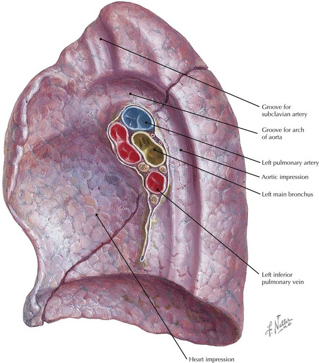
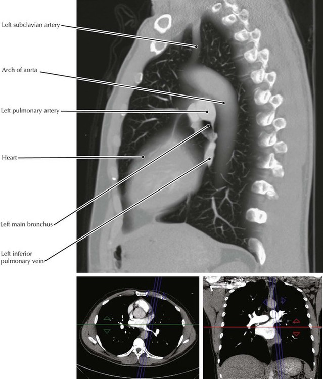
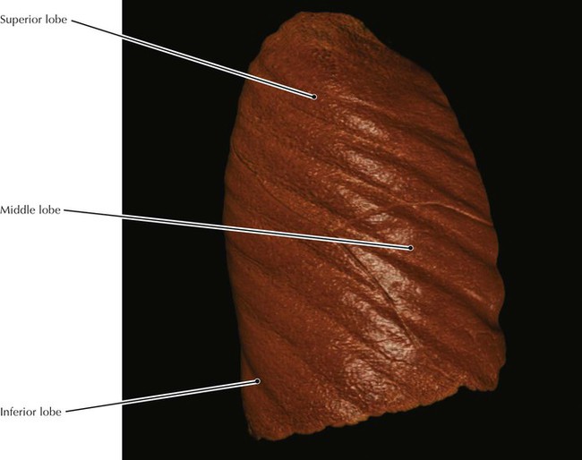
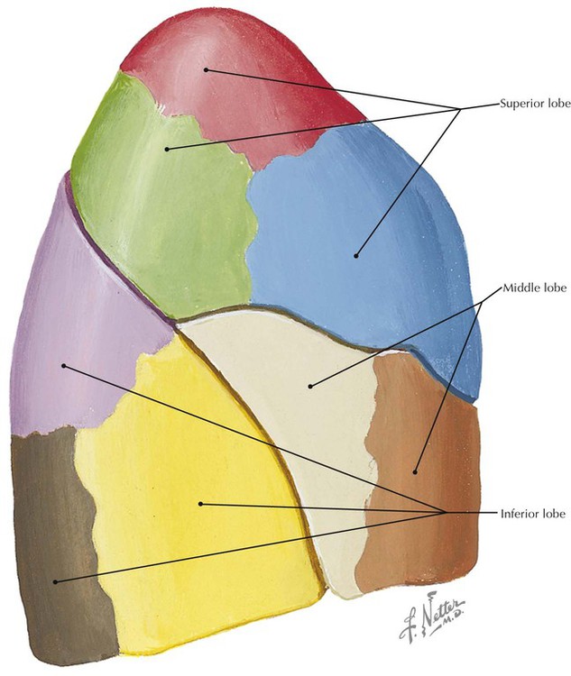
Right Lung, Lateral View
Thorax

