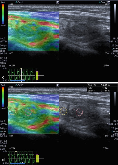
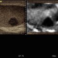
Thyroid Gland

A339375_1_En_7_Fig1b_HTML.gifA339375_1_En_7_Fig1c_HTML.jpg


Fig. 7.1
Hyperplastic follicular lesion histologically proven. (a) At baseline ultrasound the lesion appears iso-hypoechoic with peripheral halo sign. (b1, b2) At power Doppler and at Advanced Dynamic Flow™ (ADF), the nodule shows pattern III. (c) Qualitative evaluation with SE shows score 1 by Itoh et al. and Rubaltelli et al. (d) At semiquantitative evaluation, nodule appears benign with a strain ratio of 1.05 under the cutoff value of 2.0


A339375_1_En_7_Fig2_HTML.jpg


Fig. 7.2
Benign nodule at FNAC. Isoechoic nodule with halo sign which shows score 2 at qualitative evaluation


A339375_1_En_7_Fig3a_HTML.jpgA339375_1_En_7_Fig3b_HTML.jpg


Fig. 7.3
Papillary carcinoma histologically proven. (a1, a2) Hypoechoic nodule with irregular margins and pattern I at color Doppler evaluation. (b) At qualitative evaluation the nodule appears hard


A339375_1_En_7_Fig4_HTML.jpg


Fig. 7.4
Anaplastic carcinoma (histologically proven). (a) B-mode of the tumor revealed diffuse infiltration with cervical lymphadenopathy. (b) Elastography revealed stiff tissue


A339375_1_En_7_Fig5_HTML.jpg


Fig. 7.5
Papillary carcinoma histologically proven. (a) Baseline ultrasound shows an isoechoic nodule with a partially continuous halo sign and pattern III at SMI evaluation. (b) At shear wave elastography, the lesion appears hard with high elasticity values. It was a carcinoma histologically proven


A339375_1_En_7_Fig6_HTML.jpg


Fig. 7.6
Papillary carcinoma histologically proven. (a) Iso-hypoechoic nodule with high peri- and intranodular vascularization (pattern III). (b) At SSI the lesion presents mixed stiffness but high elasticity values expressed in m/s and k/Pa


References


1.

Bamber J, Cosgrove D, Dietrich CF, et al. EFSUMB guidelines and recommendations on the clinical use of ultrasound elastography. Part 1: basic principles and technology. Ultraschall Med. 2013;34(2):169–84.CrossRefPubMed


2.

Shiina T, Nightingale KR, Palmeri ML, et al. WFUMB guidelines and recommendations for clinical use of ultrasound elastography: Part 1: basic principles and terminology. Ultrasound Med Biol. 2015;41(5):1126–47.CrossRefPubMed


3.

Remonti LR, Kramer CK, Leitao CB, Pinto LC, Gross JL. Thyroid ultrasound features and risk of carcinoma: a systematic review and meta-analysis of observational studies. Thyroid. 2015;25:538–50.CrossRefPubMedPubMedCentral


4.

Brander AE, Viikinkoski VP, Nickels JI, Kivisaari LM. Importance of thyroid abnormalities detected at US screening: a 5-year follow-up. Radiology. 2000;215:801–6.CrossRefPubMed


5.

Chammas MC, Gerhard R, de Oliveira IR, Widman A, de Barros N, Durazzo M, Ferraz A, Cerri GG. Thyroid nodules: evaluation with power Doppler and duplex Doppler ultrasound. Otolaryngol Head Neck Surg. 2005;132:874–82.CrossRefPubMed


6.

Tramalloni J, Leger A, Correas JM, Monpeyssen H, Szwagier-Uzzan C, Helenon O, Moreau JF. Imaging of thyroid nodules. J Radiol. 1999;80:271–7.PubMed


7.

Reading CC, Charboneau JW, Hay ID, Sebo TJ. Sonography of thyroid nodules: a “classic pattern” diagnostic approach. Ultrasound Q. 2005;21:157–65.CrossRefPubMed


8.

Haugen BRM, Alexander EK, Bible KC, Doherty G, Mandel SJ, Nikiforov YE, Pacini F, Randolph G, Sawka A, Schlumberger M, Schuff KG, Sherman SI, Sosa JA, Steward D, Tuttle RMM, Wartofsky L. 2015 American Thyroid Association Management guidelines for adult patients with thyroid nodules and differentiated thyroid cancer. Thyroid. 2015;26(1):1–133.CrossRef


9.

Ferraioli G, Filice C, Castera L, Choi BI, Sporea I, Wilson SR, Cosgrove D, Dietrich CF, Amy D, Bamber JC, Barr R, Chou YH, Ding H, Farrokh A, Friedrich-Rust M, Hall TJ, Nakashima K, Nightingale KR, Palmeri ML, Schafer F, Shiina T, Suzuki S, Kudo M. WFUMB guidelines and recommendations for clinical use of ultrasound elastography: Part 3: liver. Ultrasound Med Biol. 2015;41:1161–79.CrossRefPubMed


10.

Tunbridge WM, Evered DC, Hall R, et al. The spectrum of thyroid disease in a community: the Whickham survey. Clin Endocrinol (Oxf). 1977;7(6):481–93.CrossRef


11.

Davies L, Welch HG. Increasing incidence of thyroid cancer in the United States, 1973–2002. JAMA. 2006;295(18):2164–7.CrossRefPubMed


12.

Sipos JA. Advances in ultrasound for the diagnosis and management of thyroid cancer. Thyroid. 2009;19:1363–72.CrossRefPubMed


13.

Iannuccilli JD, Cronan JJ, Monchik JM. Risk for malignancy of thyroid nodules as assessed by sonographic criteria: the need for biopsy. J Ultrasound Med. 2004;23(11):1455–64.PubMed


14.

Fish SA, Langer JE, Mandel SJ. Sonographic imaging of thyroid nodules and cervical lymph nodes. Endocrinol Metab Clin North Am. 2008;37(2):401–17.CrossRefPubMed


15.

Kim HG, Moon HJ, Kwak JY, Kim EK. Diagnostic accuracy of the ultrasonographic features for subcentimeter thyroid nodules suggested by the revised American Thyroid Association guidelines. Thyroid. 2013;23(12):1583–9.CrossRefPubMed


16.

Hoang JK, Lee WK, Lee M, Johnson D, Farrell S. US features of thyroid malignancy: pearls and pitfalls. Radiographics. 2007;27(3):847–60, discussion 861–865.CrossRefPubMed


17.

Moon H-G, Jung E-J, Park ST, et al. Role of ultrasonography in predicting malignancy in patients with thyroid nodules. World J Surg. 2007;31(7):1410–6.CrossRefPubMed


18.

Horvath E, Majlis S, Rossi R, Franco C, Niedmann JP, Castro A, Dominguez M. An ultrasonogram reporting system for thyroid nodules stratifying cancer risk for clinical management. J Clin Endocrinol Metab. 2009;94:1748–51.CrossRefPubMed


19.

Kwak JY, Han KH, Yoon JH, Moon HJ, Son EJ, Park SH, Jung HK, Choi JS, Kim BM, Kim EK. Thyroid imaging reporting and data system for US features of nodules: a step in establishing better stratification of cancer risk. Radiology. 2011;260:892–9.CrossRefPubMed


20.

Tee YY, Lowe AJ, Brand CA, Judson RT. Fine-needle aspiration may miss a third of all malignancy in palpable thyroid nodules: a comprehensive literature review. Ann Surg. 2007;246(5):714–20.CrossRefPubMed


21.

Peng Y, Wang HH. A meta-analysis of comparing fine-needle aspiration and frozen section for evaluating thyroid nodules. Diagn Cytopathol. 2008;36(12):916–20.CrossRefPubMed


22.

Oertel YC, Miyahara-Felipe L, Mendoza MG, Yu K. Value of repeated fine needle aspirations of the thyroid: an analysis of over ten thousand FNAs. Thyroid. 2007;17(11):1061–6.CrossRefPubMed


23.

Lyshchik A, Higashi T, Asato R, Tanaka S, Ito J, Mai J, Pellot-Barakat C, Insana M, Brill A, Saga T, Hiraoka M, Togashi K. Thyroid gland tumor diagnosis at US elastography. Radiology. 2005;237:202–11.CrossRefPubMed


24.

Ahuja AT. Lupmps and bumps in the head and neck. In: Ahuja AT, Evans R, editors. Practical head and neck ultrasound. London: Greenwich Medical Media Limited; 2000. p. 87–106.CrossRef


25.

Bhatia KS, Rasalkar DD, Lee YP, Wong KT, King AD, Yuen YH, Ahuja AT. Real-time qualitative ultrasound elastography of miscellaneous non-nodal neck masses: applications and limitations. Ultrasound Med Biol. 2010;36:1644–52.CrossRefPubMed

Only gold members can continue reading. Log In or Register to continue

Stay updated, free articles. Join our Telegram channel

Jun 4, 2017 | Posted by in ULTRASONOGRAPHY | Comments Off on Thyroid Gland

Full access? Get Clinical Tree

Get Clinical Tree app for offline access