KEY FACTS
Imaging
- •
Color, power, spectral Doppler US is screening modality for transplant renal artery stenosis
- •
Stenosis occurs most commonly at arterial anastomosis but can occur along length of artery
- •
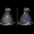
Focal elevation of peak systolic velocity (PSV) > 250-300 cm/s at stenosis with poststenotic turbulence
- •
Color aliasing and soft tissue vibration at area of stenosis
- •
Renal artery to Iliac PSV ratio > 1.8-3.5
- •
Secondary sign: Tardus parvus intrarenal waveforms = slow systolic upstroke and decreased peak velocity
- ○
Decreased acceleration index < 3 m/s² and increased acceleration time > 0.1 s in segmental arteries
- ○
Low resistive index (RI) < 0.5
- ○
- •
Catheter angiography is gold standard for diagnosis and allows treatment with angioplasty or stenting
Top Differential Diagnoses
- •
Abrupt renal artery curves and kinks
- •
Pseudorenal artery stenosis from iliac stenosis
- •
Systemic hypotension may cause low RI
Pathology
- •
Surgical injury during harvesting or transplantation
- •
Immune-mediated vascular damage from rejection
- •
Older donors and recipients, atherosclerosis, diabetes
Clinical Issues
- •
2-10% of transplants
- •
Present with hypertension, acute renal failure or progressive decline in renal function, bruit
Scanning Tips
- •
Careful attention to Doppler angle to ensure accurate PSV measurements
- •
Compare highest peak renal artery velocity to iliac artery velocity
- •
Curves and kinks may elevate velocities, mimicking stenosis
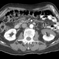
 in the main renal artery exceeds 400 cm/s with aliasing of the Doppler spectrum indicating a significant artery stenosis. Renal function was poor.
 was 151 cm/s with a renal artery to iliac artery ratio > 2.6.
Stay updated, free articles. Join our Telegram channel

Full access? Get Clinical Tree


