Trauma and Mobile Imaging
OBJECTIVES
After studying this chapter, the student will be able to:
1. List the general guidelines for radiographers to follow.
2. List members of the emergency room health care team.
3. List the basic rules in trauma radiography.
4. List common tubes and lines demonstrated with mobile chest radiographic images.
5. List the basic supplies necessary when performing trauma and mobile radiographic procedures.
6. List the precautions a radiographer should take when the patient has a head injury.
7. List the precautions a radiographer should take when the patient has facial injuries.
8. List the precautions a radiographer should take if the patient may have a spinal cord injury.
9. List the precautions a radiographer should take if the patient may have a fracture.
10. List the precautions a radiographer should take when the patient has abdominal trauma or is in acute abdominal distress.
KEY TERMS
Abrasion: A scraping or rubbing away of the surface skin by friction
Cervical: Of or pertaining to the neck
Contusion: An injury that does not break the skin; caused by a blow to the body; characterized by swelling, discoloration, and pain
Ecchymosis: An oozing of blood from a vessel into tissues, forming a discolored area on the skin
Ectopic pregnancy: An abnormal pregnancy in which the embryo implants outside the uterine cavity
Fracture: A disturbance in the continuity of a bone
Gait: Manner of moving or walking
Hemiparesis: Paralysis affecting one side of the body
Hemothorax: Collection of blood in the pleural cavity
Hypovolemic: Abnormally decreased volume of circulating fluid (plasma) in the body
Lucidity: Clearness of mind
Paresthesia: An abnormal sensation, such as burning, itching, tickling, or tingling
Pneumothorax: Accumulation of air or gas in the pleural cavity, resulting in collapse of the lung on the affected side
Somatic: Pertaining to or characteristic of the body (soma)
Mobile or portable imaging procedures are performed on patients who cannot be transported to the imaging department because of a serious injury, illness, or condition. Therefore, many patients are imaged with the use of mobile radiography equipment in the emergency department; in intensive care, coronary care, neonatal intensive care units; in special care rooms; in the patient’s room; or in the operating rooms (Fig. 10-1).
The trauma patient may be strapped to a backboard with a cervical collar and splints in place. These patients commonly have oxygen, intravenous (IV) tubing, and life support equipment when the radiographer is to perform trauma or mobile radiography procedures. Because these patients cannot be transported to radiology, the radiographer must adapt his skills to achieve diagnostic images according to the patient’s condition and needs. Specifically, a radiographer must adapt positioning and technical considerations (the central ray, image receptor, and exposure factors) during the course of performing trauma and mobile radiography. In addition, the radiographer
must analyze each patient situation in relation to the procedures requested by the physician, while at the same time keeping in mind general radiation protection measures. Preplanning is essential for achieving diagnostic outcome images under trauma or mobile circumstances. This chapter introduces additional information that is needed in trauma and mobile radiography.
must analyze each patient situation in relation to the procedures requested by the physician, while at the same time keeping in mind general radiation protection measures. Preplanning is essential for achieving diagnostic outcome images under trauma or mobile circumstances. This chapter introduces additional information that is needed in trauma and mobile radiography.
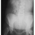
 FIGURE 10-1 (A) Setting up for a portable anterior-posterior (AP) chest semi-erect. (B) Setting up for a portable AP abdomen. (C) AP supine radiographic image of the abdomen. |
TRAUMATIC INJURIES
Traumatic injuries are caused by external force or violence (Fig. 10-2). Injury (trauma) is the leading cause of death among all persons under the age of 44. The injuries may be the result of:
Motor vehicle accidents
Pedestrian accidents
Motorcycle accidents
Falls from heights
Assaults (stabbings and gunshot wounds)
Blunt trauma
Choking
Industrial accidents
Suicides
Drowning
Smoke inhalation
Sports injuries
The trauma patient presents a wide variety of challenges for the radiographer. The nature of the injury to the patient and the patient’s condition require the radiographer to use critical thinking and judgment of a knowledgeable radiographer. Technical knowledge combined with the ability to adapt creatively is required to provide the physician(s) with the necessary diagnostic information to treat the patient. In some cases, trauma radiographs are obtained rapidly to screen for life-threatening injuries.
DISPLAY 10-1 Emergency Team Members
|
 FIGURE 10-2 Traumatic injury caused by external force. |
The radiographer is one of the first members of the health care team to see the patient once the patient is admitted to the emergency room after traumatic injury or acute illness (Display 10-1). Trauma patients can have a single injury or multiple injuries. When the radiographer is called to the emergency room to perform diagnostic radiographic images, they must assume that they will be exposed to the patient’s blood or body fluids. To avoid infecting oneself or the patient, always maintain standard precautions by having clean disposable gloves available before beginning the procedure. One must wear
the appropriate personal protective equipment that may include gloves, mask, goggles, a protective gown, and a lead or lead equivalent apron when caring for a patient who is hemorrhaging or who is nauseated and vomiting. If it is necessary to touch an open wound, wear sterile gloves. In addition, the image receptor must be protected with an impermeable covering or bag.
the appropriate personal protective equipment that may include gloves, mask, goggles, a protective gown, and a lead or lead equivalent apron when caring for a patient who is hemorrhaging or who is nauseated and vomiting. If it is necessary to touch an open wound, wear sterile gloves. In addition, the image receptor must be protected with an impermeable covering or bag.
Many patients admitted to the emergency room are in severe pain. Depending on a patient’s injuries, the radiographer will be required to assist in diagnosing the extent of injuries or illness by taking a series of images. This requires patience and skill. The radiographer will need to accomplish the procedure without extending present injuries or increasing the patient’s discomfort. Usually, time for achieving the goal is brief because the patient’s life is at risk. In trauma radiography, general radiation safety measures must be combined with speed and accuracy.
If the injured or acutely ill patient is transferred from the emergency room to the diagnostic imaging department for images, the radiographer must observe the patient for symptoms of shock. The radiographer assesses the patient’s neurologic status and level of consciousness before beginning any procedure, and then reassesses every 5 to 10 minutes while the patient is in the department. If any changes in the patient’s condition are observed, the physician and the emergency team are quickly alerted, and the radiographer prepares to assist with emergency measures, as outlined in Chapter 9.
General Guidelines
The following are the general guidelines when caring for a patient who has traumatic injuries:
1. Do not remove dressings or splints.
2. Do not move patients who are on a stretcher or backboard until ordered to do so by the physician in charge of the patient.
DISPLAY 10-2 Helpful Tips for Equipment and Accessories Needed
Lead aprons and other protective apparel for the radiographer and others involved in the procedure
Radiation detection monitoring dosimeter
Universal precaution supplies (gloves, gown, mask as needed)
Impermeable image receptor (cassette covers)
Image receptors and grids (if required)
Right or left markers and other markers to indicate whether x-ray beam is perpendicular or angled to the image receptor
Mobile (portable) or C-arm unit
3. When performing an initial cross-table lateral cervical spine radiograph, never move the patient’s head or neck or remove the cervical collar. The physician must interpret the radiograph and “clear” the cervical spine for injury before removing the collar or moving the patient.
4. Request direction from the emergency room team when planning moves, and assemble adequate assistance to move the patient safely and as painlessly as possible.
5. Do not disturb impaled objects. Support them so that they do not move as you image the patient.
6. Do not remove pneumatic antishock garments.
7. Have oxygen, suction equipment, and an emesis basin ready for use.
8. Work quickly, efficiently, and accurately to minimize repeat images.
Basic Rules for Trauma Radiography
The following are the basic rules for trauma radiography:
Assess the situation and develop an action plan for the imaging procedure.
Verify the patient’s identification with two patient identifiers.
Verify the x-ray examination with the physicians order.
Introduce yourself to the patient.
Determine patient mobility, and explain the procedure to the patient.
Predetermine equipment and accessories needed for the procedure (Display 10-2).
Announce “x-ray” before making an exposure to allow personnel to leave the area for mobile procedures.
Take at least two radiographs at 90° to one another for each body part (Fig. 10-3).

FIGURE 10-3 (A) Trauma lateral right ankle. (B) Second projection 90° from lateral ankle.
Make sure that the central ray and image receptor alignment approaches routine positioning applications, adapting to the patient’s condition.
Include all anatomy of interest.
For long-bone radiography, include both the joints; two exposures may be required with overlap (1.5 to 2 inches) to ensure that the entire bone is included.
Provide radiation safety protective apparel for anyone who needs to be in the room caring for the injured patient.
A cooperative environment with all personnel involved in the emergency procedure will facilitate the proper care of the patient.
THE PATIENT WITH A HEAD INJURY
Injuries to the head are exceedingly common. Each year approximately 10 million head injuries are predicted to occur in the United States. The term “head injury” may refer to any injury of the skull, brain, or both that requires medical attention. Radiographers are not called as often as in past years to perform radiographic images because of the availability and effectiveness of computed tomography (CT), which allows the physicians to diagnose head injuries (Fig. 10-4). All head injuries are potentially serious because they may involve the brain, which is the seat of consciousness and controls every human action.
The two basic types of head injury are open and closed. With an open injury to the skull or meninges, the brain is vulnerable to damage and infection because its protective casing has been broken. If the injury is closed
(also called a blunt injury), the brain tissue may swell. The swelling is limited by the confines of the skull, and the resulting pressure may cause extensive brain damage. The brain has little healing power, so any injury to it must be considered potentially permanent and serious.
(also called a blunt injury), the brain tissue may swell. The swelling is limited by the confines of the skull, and the resulting pressure may cause extensive brain damage. The brain has little healing power, so any injury to it must be considered potentially permanent and serious.
 FIGURE 10-4 CT image for head trauma. |
Fractures at the base of the skull (basal skull fractures) often have accompanying fractures of the facial bones. This type of injury may result in a tear in the dura mater, the outer membrane surrounding the brain and spinal cord, and a leakage of the cerebrospinal fluid (CSF) may result.
Consider all patients with head injuries to have accompanying cervical spinal injuries until it is medically disproved. Take precautions to alleviate potential extension of these injuries as a matter of routine. It is vital for the radiographer to be knowledgeable in understanding the signs and symptoms of patients with a variety of injuries. It is important to report any observable changes in the patient’s status to the emergency physician and/or team.
Clinical Manifestations of Head Injury
Closed Injury
Varying levels of consciousness, ranging from drowsiness, confusion, irritability, and stupor to coma
Lucid periods followed by periods of unconsciousness are possible
Loss of reflexes
Changes in vital signs
Headache, visual disturbances, dizziness, and giddiness
Gait abnormalities
Unequal pupil dilation
Seizures, vomiting, and hemiparesis
Open Injury
Abrasions, contusions, or lacerations apparent on the skull
A break or penetration in the skull or meninges apparent by inspection or on radiographic images
Basal fractures resulting in leakage of CSF, as demonstrated by leakage of blood on the sheet or dressing surrounded by a yellowish stain (the halo sign); CSF may leak from the nose or ears or as a postnasal drip
Varying levels of consciousness
Subconjunctival hemorrhage
Hearing loss
Periorbital ecchymosis (raccoon eyes)
Facial nerve play
Radiographer’s Response
1. Keep the patient’s head and neck immobilized until the physician rules out injury to the spinal cord.
2. If possible, elevate the patient’s head 15° to 30°.
3. Do not remove sandbags, collars, or dressings. Take all radiographic images with these in place.
4. Do not flex the patient’s neck or turn it to either side. Rotation of the head may increase intracranial pressure.
5. Keep the patient’s body temperature as normal as possible. Do not allow the patient to become chilled or overheated.
Related posts:


Stay updated, free articles. Join our Telegram channel

Full access? Get Clinical Tree


