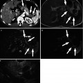
Fig. 16.1
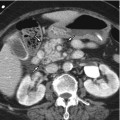
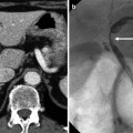
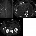
Traumatic injury of bile duct. (a) Penetrating injury of segment 4 of the liver by the iron bar (arrow) in a construction worker. A pseudoaneurysm (arrowhead) of the hepatic artery is seen in the liver hilum. (b) Follow-up CT scan 2 months later from the initial CT scan. Bilomas (arrows) are seen in perihepatic and subhepatic spaces. (c) ERCP image shows the leakage of the contrast medium (white arrow) from the bile duct to one of the biloma (black arrow). Coils (arrowheads) used for the embolization of pseudoaneurysm are seen
16.5.2 Strasberg Classification of Bile Duct Injury
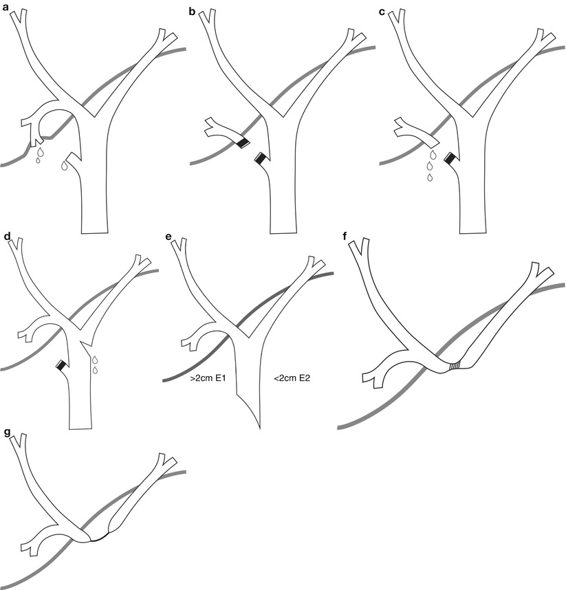
Fig. 16.2
Strasberg classification of bile duct injury. (a) Class A. Bile leak from the cystic duct or liver bed. (b) Class B. Occlusion of part of the biliary tree, typically an aberrant segmental duct or sectoral duct. (c) Class C. Bile leak from an aberrant segmental duct or sectoral duct. (d) Class D. Partial lateral injury of the extrahepatic bile duct. (e) Class E1 and E2. (E1) Circumferential injury of the common hepatic duct with a residual stump longer than 2 cm. (E2) Circumferential injury of the common hepatic duct with a residual stump shorter than 2 cm. (f) Class E3. Hilar injury at the confluence with preservation of hepatic duct continuity. (g) Class E4. Hilar injury at the confluence with loss of hepatic duct continuity
16.5.3 Normal Cholangiography After Cholecystectomy
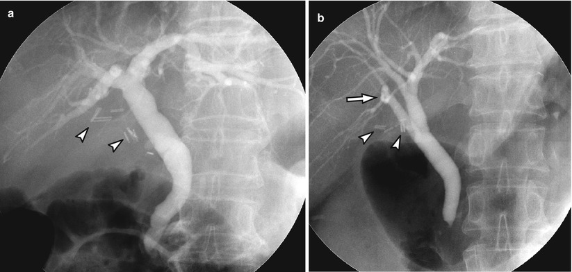
Fig. 16.3
Normal cholangiography after cholecystectomy. (a) Cholangiography of bile duct with normal anatomy. Surgical clips (arrowheads) are seen at the site of cholecystectomy. (b) Cholangiography of bile duct with anatomical variation. Right posterior duct (arrow) drains directly to the common hepatic duct. Surgical clips (arrowheads) are seen at the site of cholecystectomy
16.5.4 Postcholecystectomy Bile Duct Injury. Class A
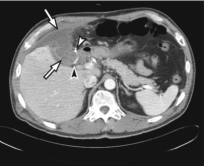
Fig. 16.4
Postcholecystectomy bile duct injury. Class A. Bile collection in gallbladder fossa and perihepatic space (arrows). A surgical clip (white arrowhead) and a surgical drain (black arrowhead) are seen around the bile collection
16.5.5 Postcholecystectomy Bile Duct Injury. Class B
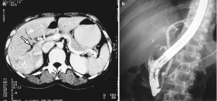
Fig. 16.5
Postcholecystectomy bile duct injury. Class B. (a) Dilatation of the aberrant right posterior inferior segmental duct (B6, arrow). (b) On ERCP common duct and left segmental ducts are seen. Isolated right posterior inferior duct (B6) is not opacified. In addition, right anterior segmental duct is not seen because of postoperative stricture
16.5.6 Postcholecystectomy Bile Duct Injury. Class B
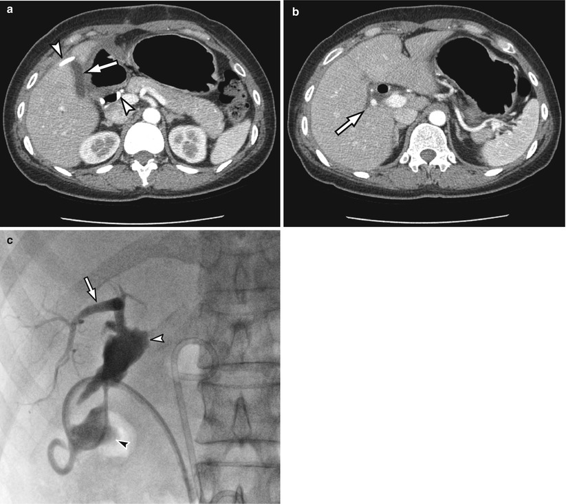
Fig. 16.6
Postcholecystectomy bile duct injury. Class B. (a) A small biloma (arrow) is seen in the gallbladder fossa. A drainage catheter (arrowheads) is inserted to remove bile collection in subhepatic space. (b) Right posterior inferior segmental duct (B6, arrow) is dilated because of the incidental ligation during the operation. (c) After injection of contrast medium through the catheter, biloma in the subhepatic space (black arrowhead), gallbladder fossa (white arrowhead), and B6 duct (arrow) are visualized
16.5.7 Postcholecystectomy Bile Duct Injury. Class D
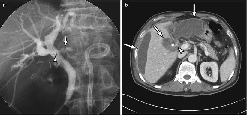
Fig. 16.7
Postcholecystectomy bile duct injury. Class D. (a) A T-tube (arrowhead) was inserted at the site of common hepatic duct injury during the laparoscopic cholecystectomy. On ERCP examination, contrast medium (arrow) flows out of the bile duct through the injury site. (b) On CT scan, bilomas (arrows) are seen in perihepatic and subhepatic spaces and cholecystectomy site with surgical clips (arrowhead)
16.5.8 Postcholecystectomy Bile Duct Injury. Class E2
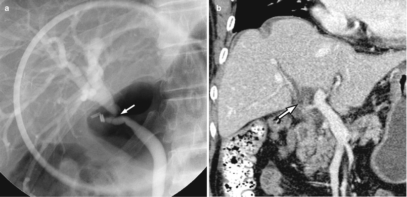
Fig. 16.8
Postcholecystectomy bile duct injury. Class E2. Abrupt narrowing of common hepatic duct (arrow) with short (<2 cm) stump on ERCP (a) and CT (b)
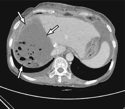
16.5.9 Biloma After Right Hemihepatectomy

