Traumatic Bronchial Injury
Sam A. Glaubiger
CLINICAL HISTORY
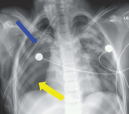
22-year-old male after high-speed motor vehicle collision with continuous air leak from right chest tube and lack of right lung reinflation.
FINDINGS
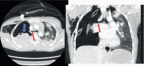
Figure 18A: Anteroposterior (AP) supine film of the chest. Right chest tube (blue arrow) is in place. No normal right lung markings are present, and lung is not inflated (yellow arrow). Figure 18B: Axial contrast-enhanced CT image of the chest. The right lung (blue arrow) is completely collapsed, and has “fallen” into the dependent right hemithorax. Air fills the remainder of the right hemithorax. A right mainstem bronchial stump (red arrow) confirms complete bronchial laceration. Left-sided opacity is a pulmonary contusion.
DIFFERENTIAL DIAGNOSIS
Bronchial injury, esophageal injury, pulmonary laceration.
DIAGNOSIS
Bronchial injury.
