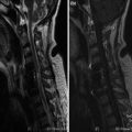
Fig. 1
a–f. SE T1 (a–b), FSE T2 (c–d), STIR (e–f) sagittal sections
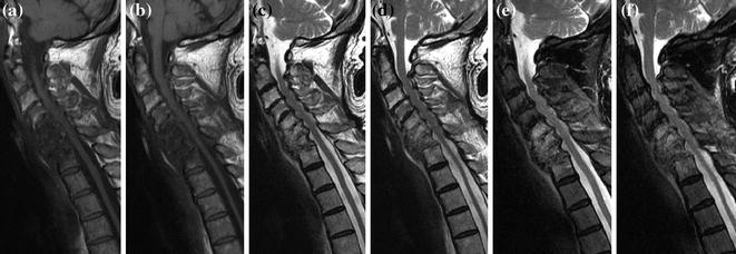
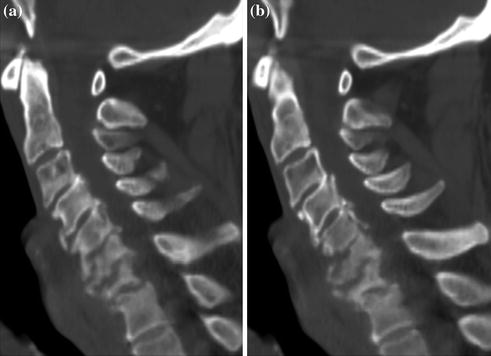
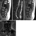
Fig. 2
a–f. SE T1 (a–b), FSE T2 (c–d), STIR (e–f) sagittal sections. Reabsorption of epidural hematoma at C7–D1; C6–C7 eight reduction and anterior wedging. C5–C7 edema
Only gold members can continue reading. Log In or Register to continue
Related posts:
Stay updated, free articles. Join our Telegram channel

Full access? Get Clinical Tree


