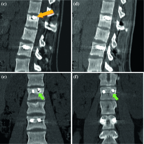
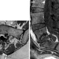
Fig. 1
a–f. CT D12-L4 (a, b), MPR sagittal (c, d) and coronal (e, f). Bone alteration surrounding left L1 transpedicular screw with rarefaction and peripheral sclerosis (a, b, e, f, short arrow); front end screw reaches L1 superior surface (c, long arrow). L2 fracture with superior surface depression (e)
< div class='tao-gold-member'>
Only gold members can continue reading. Log In or Register to continue
Related posts:
Stay updated, free articles. Join our Telegram channel

Full access? Get Clinical Tree


