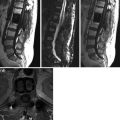
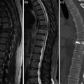
Fig. 1
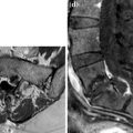
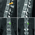
a–b. CT thorax-abdomen a, MPR sagittal b. L2–L3 irregular alignment
59.2
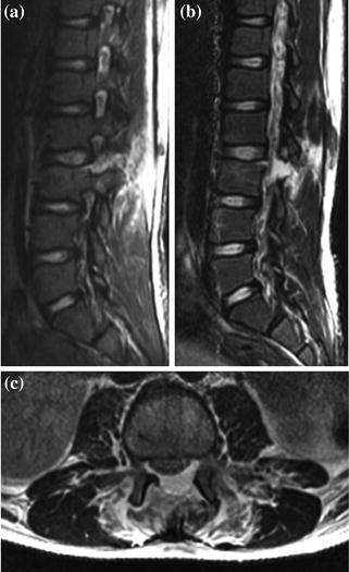
Fig. 2
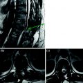
a–c. STIR sagittal (a), FSE fat sat T2 sagittal (b) and axial (c). Interspinous L2–L3 gap and opening of interapophyseal articulations in traumatic hyper-flexion. Complete rupture of L2–L3 yellow and interspinous ligaments. Posterior L2–L3 disk detachment from L3 with probably rupture of posterior longitudinal ligament
< div class='tao-gold-member'>
Only gold members can continue reading. Log In or Register to continue
Stay updated, free articles. Join our Telegram channel

Full access? Get Clinical Tree


