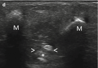
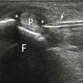
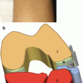
Fig. 19.1


US-guided treatment of Morton’s neuroma with coaxial approach. (a) Probe and patient position to perform US-guided treatment of Morton’s neuroma. (b) Anatomical scheme and (c) US image of perineural needle insertion, M metatarsal heads, T flexor tendons, S subcutaneous tissue, arrow needle tip, arrowheads neuroma, B intermetatarsal bursa. (d) Steroid injection (asterisk)
Related posts:


Stay updated, free articles. Join our Telegram channel
Full access? Get Clinical Tree
