| SKULL BASE REGION | Meckel’s cave and posterior cranial fossa |
| HISTOPATHOLOGY | N/A |
| PRIOR SURGICAL RESECTION | No |
| PERTINENT LABORATORY FINDINGS | N/A |
Case description
A 70-year-old male patient with known history of a lesion compatible with a trigeminal schwannoma (TS) presented to our care 8 years after initial imaging with right V2 hypesthesia. Repeat magnetic resonance imaging (MRI) revealed a major volumetric increase of the TS ( Figure 5.21.1 ). A microsurgical resection was proposed, but the patient refused surgery. Due to the large volume of the lesion, as well as compression of the brainstem with deformation of the fourth ventricle, a staged-volume Gamma Knife radiosurgery (GKR) was performed ( Figure 5.21.2 ).
| Radiosurgery Machine | Gamma Knife – Icon |
| Radiosurgery Dose (Gy) |
|
| Biologically Effective Dose (Gy) |
|
| Number of Fractions | 1 for each stage |

T1-gadolinium injected MRI in the axial plane displaying a large lesion, compatible with a TS, with perilesional edema and displacement of the fourth ventricle without hydrocephalus. TS, Trigeminal schwannoma.

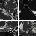
From left to right and up to down: multimodal radiosurgery therapeutic imaging including T1 noninjected, T1-gadolinium injected, T2 CISS/Fiesta noninjected, and T2 CISS/Fiesta injected MRI. The initial dosimetry (stage 1) is colored in blue and superimposed on the subsequent dosimetry (stage 2, colored in yellow), and was performed 11 months later. Central loss of enhancement is noted within the first stage (blue line, after 11 months, at the time of the second GKR). There was no hydrocephalus. Perilesional edema was stable. The marginal dose was 12 Gy for each stage. CISS, Constructive interference in steady state; GKR, Gamma Knife radiosurgery.
| Critical Structure | Dose Tolerance |
|---|---|
| Brainstem | Marginal dose is 12 Gy, although there is contact between TS and the brainstem; the risk of adverse radiation events (ARE) at the brainstem level remains virtually zero due to such a low marginal dose prescription |
| Cochlea | Maximal dose is 1.9 Gy, or 1.4 Gy (for volume-staged treatment); safe maximal dose to cochlea for hearing preservation can be as high as 4 to 5 Gy |
| Optic chiasm | Maximal dose is 1.4 Gy; safe maximal dose to optic apparatus for optic pathway preservation can be as high as 12 and 14 Gy |
Related posts:
 Esthesioneuroblastoma – delayed postoperative radiosurgery for recurrence at long-term
 Null cell – delayed postoperative radiosurgery for growing perioptic residual
 Chordoma – immediate postoperative/post-proton therapy radiosurgery for residual disease
 Trigeminal neuralgia due to microvascular conflict – upfront radiosurgery
 Capillary hemangioma – postoperative radiosurgery for residual tumor
 Superior sagittal sinus meningioma – delayed postoperative, multisession radiosurgery for growing residual
Stay updated, free articles. Join our Telegram channel

Full access? Get Clinical Tree


