| SKULL BASE REGION | Suprasellar/tuberculum sella |
| HISTOPATHOLOGY | N/A |
| PRIOR SURGICAL RESECTION | No |
| PERTINENT LABORATORY FINDINGS | N/A |
Case description
The patient is a 54-year-old female who was found to have a mild left superior nasal field cut on her annual visual examination. Brain magnetic resonance imaging (MRI) revealed a tuberculum sella meningioma causing mild mass effect on the optic tract/chiasm ( Figure 4.17.1 ). Since the patient did not have any subjective visual field deficits, stereotactic radiosurgery (SRS) was performed ( Figure 4.17.2 ).
| Radiosurgery Machine | CyberKnife |
| Radiosurgery Dose (Gy) | 25, at the 80% isodose line |
| Number of Fractions | 5 |

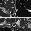
Postcontrast axial, sagittal, and coronal images showing a 1.1 × 1.4 × 0.9 cm homogeneously enhancing mass in the suprasellar region exactly right of the midline, causing a mass effect on the optic tract/chiasm.

Imaging of the treatment plan.
| Critical Structure | Dose Tolerance |
|---|---|
| Optic nerve/chiasm | 25 max per 5 fractions / <0.2cc>20 Gy |
| Brainstem |
|
| Cranial nerves in cavernous sinus |
|
| Cavernous carotid artery |
|
Related posts:
 Esthesioneuroblastoma – delayed postoperative radiosurgery for recurrence at long-term
 Null cell – delayed postoperative radiosurgery for growing perioptic residual
 Chordoma – immediate postoperative/post-proton therapy radiosurgery for residual disease
 Trigeminal neuralgia due to microvascular conflict – upfront radiosurgery
 Capillary hemangioma – postoperative radiosurgery for residual tumor
 Superior sagittal sinus meningioma – delayed postoperative, multisession radiosurgery for growing residual
Stay updated, free articles. Join our Telegram channel

Full access? Get Clinical Tree


