Classification
WHO grade
Astrocytic a
Pilocytic
I
Diffuse
II
Anaplastic
III
Glioblastoma
IV
Oligodendroglial and oligoastrocytic a
Oligodendroglioma
II
Oligoastrocytoma
II
Anaplastic oligodendroglioma
III
Anaplastic oligoastrocytoma
III
Glioblastoma with oligodendroglioma component
IV
Ependymal a
I–III
Choroid plexus
I, III
Neuronal and mixed neuronal-glial
I–II
Pineal parenchymal
II, IV
Embryonal
IV
Meningeal
I–III
1 Biomarkers
Diagnosis and treatment of glioma is rapidly shifting away from a purely histopathologic paradigm towards one that incorporates and may ultimately be replaced by biomarkers for diagnosis, prognosis, and selection of therapy. Indeed while the current buzzwords “personalized medicine” belie a long medical tradition to seek better, more out understanding of targeted treatments, biotechnology has advanced sufficiently that molecular medicine has become considerably more granular. We can correlate new biomarkers with previously known imaging features, histopathologic findings, and patient outcomes. We may to determine if a marker is diagnostic, prognostic and/or predictive to available therapies. Often because we know long standing therapies based on older histopathologic classification empirically work, ethical consideration make it difficult to establish how newly discovered biomarkers might alter therapeutic approach. We identify an interesting genetic marker but remain in the dark with regard to what protein it codes for and wonder if it is a potential molecular target of therapy. For these reasons the current study of glioma is fascinating and frustrating (Tables 2, 3).
Table 2
Prognostic biomarkers in glioma
D× | A | Oligo | OA | AA | AO | AOA | GBM | |
|---|---|---|---|---|---|---|---|---|
1p/19q | + | NA | +a | +a | NA | + | + | NA |
MGMT | − | ? | ? | ? | + | + | + | + |
IDH | + | + | + | + | + | + | + | +a |
Table 3
Predictive biomarkers in glioma
A | O | Mixed | AA | AO | AOA | GBM | |
|---|---|---|---|---|---|---|---|
1p/19q | NA | +a | +a | NA | + | + | NA |
MGMT | ? | ? | ? | ? | ? | ? | +a |
IDH | ? | ? | ? | ? | ? | ? | ? |
1.1 1p/19 q
Co-deletion of chromosomal arms 1p and 19q is a genetic signature found in approximately 65 % of high-grade and 85 % of low-grade oligodendrogliomas (LGG). It is less commonly observed in mixed glioma or astrocytomas. The higher incidence of co-deletion in low grade glioma may suggest an early event in tumor formation (Smith et al. 2000; Barbashina et al. 2005). While histopathological interpretation is still necessary for subtyping of glial tumor, 1p/19q co-deletion can be used to support a diagnosis of oligodendroglioma where histology is ambiguous or in the event of inter-observer disagreement (Aldape et al. 2007). While the role of the gene product is undetermined, co-deletion in anaplastic oligodendroglioma is both predictive and prognostic; that is, co-deleted tumors are more responsive to chemo- and radiotherapy and have a more favorable prognosis regardless of treatment than lesions without co-deletion (Robertson et al. 2011; Hofer and Lassman 2010; Cairncross et al. 1998; Bauman et al. 2000; Wick et al. 2009b, Intergroup Radiation Therapy Oncology Group Trial 9402 et al. 2006; van den Bent et al. 2013).
Data regarding the value of 1p/19q in low-grade glioma is beginning to emerge but is not definitive. Some suggest that in the absence of adjuvant cytoxic therapy, 1p/19q status loses its prognostic impact for LGG while neuro-radiologic monitoring studies suggest a slower natural growth rate for untreated co-deleted lesions. (Robertson et al. 2011; Weller et al. 2009; Ricard et al. 2007). The Clinical Cooperation Unit Neuropathology in Germany evaluated markers including 1p/19q deletion and their relationship to progression free survival (PFS) in LGG cohorts who either did or did not receive adjuvant radio- or chemotherapy. In this study no biomarker was prognostic for PFS in the absence of adjuvant therapy while 1p/19q co-deletion was associated with both PFS and overall survival (OS) for patients receiving either adjuvant chemotherapy or radiotherapy (Smith et al. 2000; Hartmann et al. 2011; Barbashina et al. 2005). This report is interesting but limited as a retrospective study of relatively small numbers.
The impact of a single arm deletion is not definitively understood. Preliminary data from EORTC 22033–26033 trial was presented at ASCO in 2013. High-risk low-grade glioma patients were randomized to radiotherapy versus temozolomide (TMZ) and were stratified by 1p status. 1p deletion was confirmed to be a positive prognostic factor regardless of treatment. Patients with intact 1p treated with TMZ were observed to have a non-significant trend towards inferior progression-free survival. Overall survival may be superior with 1p deletion (Aldape et al. 2007; Baumert et al. 2013). However maturation of the data and analysis of 1p/19q co-deletion is required before any definitive assessment of response to therapy by these biomarkers. Results are pending from E3F05 in which patients were receiving radiotherapy ± TMZ for LGG. The study will include an analysis of 1p/19q status as it correlates to outcome (NCT 00978458).
1.2 MGMT
O6-methylguanylmethyltransferace (MGMT) is a DNA repair protein that removes alkyl groups from the O6 position of guanine. The mechanism of the anti-tumor activity of temozolomide (TMZ) is believed to be through tumor DNA-alkylation most frequently at the N7 and O6 position of the guanine moiety. Epigenetic silencing of MGMT by promoter methylation (MGMT-met) is associated with a loss of enzymatic expression and subsequent diminished DNA repair activity. Therefore tumor in which MGMT is silenced (i.e. methylated) is expected to be more sensitive to TMZ than unmethylated lesions (MGMT-unmet).
The powerful predictive and prognostic role of MGMT promoter methylation in glioblastoma was illustrated in a classic EORTC-NCIC study of GBM patients treated with radiation with and without concomitant and adjuvant temozolomide (Stupp et al. 2005; Hegi et al. 2005). In that study, patients whose tumors were MGMT methylated had a more favorable overall survival than those whose tumors were MGMT unmethylated. This finding could signify a more favorable natural outcome for MGMT-met GBM, or reflect a more favorable response to both RT and TMZ than MGMT-unmet. An analysis was not performed comparing outcomes by methylation status for patients receiving radiotherapy only. Examination of the survival curves for patients receiving RT alone demonstrates an appreciable separation by MGMT methylation status. When both treatment assignment and MGMT methylation status were considered, the most favorable outcome was found in MGMT-met patients receiving combined therapy. It is interesting to note that OS trended towards significance for combined therapy even when tumors were MGMT-unmet, arguing that alkylation of the O6 moiety on the MGMT promoter is not the sole mediator of TMZ efficacy (Hegi et al. 2005).
Because temozolomide was frequently used for salvage after progression in the radiotherapy only arm, the authors looked at the interaction of treatment assignment and MGMT status on progression-free survival to better assess the predictive nature of MGMT methylation to therapy. For patients with MGMT-met tumors, PFS was superior for patients receiving combined therapy than compared to those receiving radiotherapy alone. PFS with combined therapy was also superior for patients with MGMT-unmet tumors again suggesting alternate pathways for the efficacy of temozolomide. This study could not address whether MGMT status predicts response to RT alone (Hegi et al. 2005).
The NOA-04 trial prospectively randomized patients with grade III glioma- anaplastic oligodendroglioma (AO), anaplastic oligoastrocytoma (AOA) and anaplastic astrocytoma (AA)- to RT versus either a combination of procarbazine, lomustine and vincristine (PCV) or TMZ chemotherapy. MGMT promoter methylation was associated with PFS for both chemotherapy and radiotherapy confirming its prognostic relevance. However the trial did not support the hypothesis that MGMT methylation simply predicted response to alkylating chemotherapy (Wick et al. 2009b). Unlike the EORTC-NCIC trial of GBM, an analysis of MGMT status in the radiotherapy alone arm was performed. MGMT methylation conferred a clear and significant PFS benefit for patients receiving RT alone. The authors concluded that in anaplastic glioma, MGMT promoter hypermethylation was (1) a prognostic marker for patients treated with RT or (2) a predictive for response to RT itself (Wick et al. 2009b). The EORTC 26951 study of anaplastic oligodendroglial tumors makes for an interesting companion study. EORTC 26951 randomly assigned patients to either RT or RT followed by adjuvant PCV. MGMT promoter methylation was prognostic for both PFS and OS in both arms and did not have predictive significance for response to PCV. Interestingly in lesions identified as GBM on central review in this study no prognostic role for MGMT methylation was observed (van den Bent et al. 2013).
The Methusalem trial demonstrated that for the elderly with malignant astrocytoma (AA or GBM), MGMT-met is associated with improved OS. In the event of promoter methylation, event-free survival is improved for patients assigned to TMZ compared to those who receive RT. In the absence of methylation however, event free survival was superior for patients assigned to the radiotherapy group. There was no arm of patients receiving combined therapy (Wick et al. 2012).
The data is conflicted with regard to the value of MGMT in the prediction of response to therapy and prognostication in low-grade glioma. An association of MGMT promoter methylation with overall survival has been demonstrated. Correlation of MGMT-met with the putative prognostic marker 1p/19q co-deletion has been postulated as an explanation for this finding (Kesari et al. 2009; Leu et al. 2013). Other studies however contradict these observations (Tosoni et al. 2008). There is a paucity of data to suggest a predictive relationship between methylation status of MGMT and response to TMZ (Hartmann et al. 2011; Kesari et al. 2009; Groenendijk et al. 2011).
1.3 IDH1/IDH2
Parsons et al. used high-density oligonucleotide DNA array to detect the presence of amplifications and deletions among 20,661 coding genes in GBM samples. Recurrent mutations at codon 132 in the active site of isocitrate dehydrogenase 1 (IDH1) were noted with an incidence of 12 % (Parsons et al. 2008). The mutation was associated with younger patients, secondary GBM and improved survival. This may explain long observed association between younger age at diagnosis and improved outcome in glioma (Scott et al. 1998). Subsequent work has identified an association between IDH mutations and grade II and III astrocytoma, oligodendroglioma and oligoastrocytoma, and has identified IDH2 (codon 172) which is primarily associated with oligodendroglioma (Yan et al. 2009; Hartmann et al. 2009). IDH mutations are quite rare in de novo GBM and absent in pilocytic astrocytoma.
IDH mutations are tightly correlated with other biomarkers and histologic subtype. For instance IDH mutation is associated with 1p/19q co-deletion in oligodendroglial tumor and TP53 in astrocytoma, but seems to be mutually exclusive with EGFR and PTEN abnormalities. Combined IDH1 and IDH2 mutation is rare (Yan et al. 2009; Gupta and Salunke 2012; Sanson et al. 2009). It has been observed that IDH remains an independent favorable prognostic marker after adjustment for grade and MGMT status (Sanson et al. 2009).
In the cell IDH catalyzes oxidative decarboxylation of isocitrate to α-ketoglutarate (α-KG) in the citric acid cycle. The enzyme products of IDH1/2 utilize NADP +, which is transformed into NADPH with the generation of α-KG. These products normally protect against oxidative damage (Gupta and Salunke 2012). Both “loss of function” (production of α-KG and NADPH) and “gain of function” (accumulation of D-2-hydroxyglutarate (D2HG)) hypotheses have been put forwards to explain the mechanisms by which IDH mutation mediates oncogenesis. IDH1 mutations are tightly associated with CpG island methylator phenotype across tumor grade, raising the hypothesis that mutation may predispose cells to large-scale epigenetic disruption (Gupta and Salunke 2012; Noushmehr et al. 2010).
IDH has meaningful diagnostic potential and can be used in the clinic to increase confidence in differentiating between grade II glioma and IDH-wild type lesions such as pilocytic astrocytoma, pleomorphic xanthroastrocytoma or medulloblastoma. An IDH mutant oligodendroglial tumor may be distinguished from neurocytoma or dysembryoplastic neuroepithelial tumors and secondary GBM from de novo GBM (Gupta et al. 2011; Yan et al. 2009; Capper et al. 2011).
IDH mutation has been associated with better overall prognosis for glioma across multiple studies among several subtypes. IDH appears to have prognostic value in multivariate analysis even when controlling for other favorable variables such as low grade, young age, 1p/19q co-deletion and MGMT promoter methylation (Metellus et al. 2010; Alexander and Mehta 2011; Parsons et al. 2008; Sanson et al. 2009; Wick et al. 2009b; van den Bent et al. 2010; Weller et al. 2009; Houillier et al. 2010). IDH mutation status may have more powerful prognostic value in high-grade glioma than standard histopathologic classification. An analysis of NOA-04 demonstrated that IDH mutation status may override grade in malignant glioma in terms of prognostication (Hartmann et al. 2010; Alexander and Mehta 2011).
While several studies have demonstrated an association between mutated IDH and improved survival in glioma patients, there is as of yet no robust evidence that an IDH mutation predicts response to any therapy (Yan et al. 2009; Parsons et al. 2008; van den Bent et al. 2010; Hartmann et al. 2010). In a clinical study IDH mutation has been associated with improved response to TMZ, however this observation may be due to its association with 1p/19q (Houillier et al. 2010).
1.4 Imaging biomarkers
Imaging biomarkers (an evolution of what was previously called “Roentgen signs” in prior generations) can be useful in the diagnosis and grading of glioma however as yet cannot be considered pathognomonic for a particular pathologic sub-type of glioma or grade. Nevertheless a constellation of imaging features may direct diagnostic assessment and subsequent management. The standardization of biomarkers is evolving and their utility is appreciated. As such a rigorous criteria for imaging biomarkers as surrogate endpoints has been developed for use in clinical trials (Schatzkin and Gail 2002).
1.
The presence of the biomarker is closely coupled or linked to the condition.
2.
The detection and/or quantitative measurement of the biomarker is accurate, reproducible and feasible over time.
3.
The measured changes over time in the imaging biomarker are closely coupled or linked to the success or failure of the therapeutic effect and the true end-point sought for the medical therapy being evaluated.
Low-grade glioma tends to appear bright on magnetic resonance imaging (MRI) T2/Flair imaging and hypodense on T1-weighted images and typically do not enhance with the addition of IV contrast. An exception is grade 1 pilocytic astrocytoma which usually presents in the posterior fossa and enhances with contrast (Lee et al. 1989). The presence of even trace or wispy amounts of enhancement on T1 weighted images should raise suspicion for an at least partially high-grade lesion. If definitive resection cannot be achieved the surgeon should attempt biopsy of the enhancing region if possible in order to establish the highest-grade component of the lesion. Failure to indentify high-grade tumor in a region of enhancement by a small sample may be the result of sampling error and may not definitively rule out the possibility of the presence of malignant glioma (Pignatti 2002; Bauman et al. 1999).
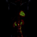
Oligodendroglial tumors have a greater propensity for the frontal and temporal lobes, and more frequently present with calcifications and hemorrhage than do astrocytomas (Lee and Van Tassel 1989). Positron-emission tomography (PET) may help to clarify clinical diagnosis. Glucose hypometabolism on PET supports a diagnosis of LGG whereas hypermetabolic activity is consistent with the presence of a high-grade lesion. Magnetic resonance spectroscopy, PET and L-(methyl-11C) methionine (MET-PET), can all contribute to direct guided biopsy of areas suspicious for malignancy (Pirotte et al. 2004; McKnight et al. 2002).
2 Low Grade Glioma
The designation low-grade glioma represents a heterogeneous population of primary tumors. For our purposes we will confine discussion to the relatively common astrocytic and oligodendroglial sub-types.
LGG often come to attention due to new on-set seizure activity (Lote et al. 1998). The acute and dramatic nature of seizure typically results in a short interval between initial symptom and diagnosis. However symptoms of LGG can often be insidious. Patients may report a vague, chronic history of headache for which they have previously received medical attention. The pain pattern is widely variable and may even remit from time to time or be at least initially responsive to conservative medical therapy. Focal neurologic deficits may develop depending upon the location of the lesion and will frequently precipitate neurologic imaging.
The gold standard for diagnosis of LGG is histo-pathologic examination of tumor. However tissue sampling may be limited by neurosurgical accessibility of the lesion and the resulting specimen non-diagnostic. Perhaps more frequently, biopsy sampling is sufficient to make a diagnosis of glioma but not to adequately grade it, the consequence of which being the potential under-treatment of a high-grade lesion. Thus in the absence of adequate tissue for full diagnosis, we rely upon other potential biomarkers as noted in the above section.
2.1 Initial Treatment. Postoperative Radiotherapy: Adjuvant or Delayed?
As there is evidence for improved survival, we recommend maximal safe resection at time of diagnosis (Smith et al. 2008; Keles et al. 2001). The role and selection of adjuvant therapy in the immediate post-operative setting is dependent upon risk factors, predictive biomarkers and neurologic status (Van den Bent et al. 2005; Pignatti 2002; Shaw 2002).
The EORTC 22845 trial assigned patients to receive RT either immediately following resection or at the time of progression. While immediate postoperative RT significantly prolonged PFS (5.4 versus 3.7 years), overall survival was not affected (7.4 versus 7.2 years) (Van den Bent et al. 2005). Importantly, this study demonstrated that malignant transformation was not more likely in patients having received post-operative radiotherapy. This study also demonstrates the efficacy of radiotherapy for treatment of low-grade glioma. Though the incidence of seizure at baseline between the two arms was comparable, at one year seizure incidence was significantly lower in patients undergoing immediate postoperative RT (25 versus 41 %) and is a justification for the use of RT for LGG patients with refractory seizure (Van den Bent et al. 2005).
Low-grade glioma patients at high risk for early and rapid progression after surgery alone have been identified. Multivariate analysis has shown that age >/= 40, pure astrocytoma subtype, diameter >/= 6 cm, tumor crossing midline and neurologic deficit prior to surgery are unfavorable prognostic features for OS. The presence of three or more high-risk features identifies patients who may warrant immediate adjuvant radiotherapy (Pignatti 2002; Shaw 2002).
2.2 Radiation Dose-Escalation
Both the EORTC and the RTOG have conducted multicenter prospective randomized trials comparing 45–50.4 Gy in conventional fraction to higher total doses of 59.4–64.8 Gy in an attempt to improve outcomes of LGG through dose-escalation. Both trials failed to demonstrate benefit and severe radionecrosis was doubled in the highest dose arm. Thus doses of 45–54 Gy remain the standard of care for low-grade glioma (Karim et al. 1996; Shaw 2002).
2.3 Adjuvant Chemotherapy?
The role of postoperative chemotherapy for LGG is still being investigated. RTOG 9802 examined patients deemed high-risk by virtue of age >/= 40 or subtotal resection. Patients were randomized to postoperative RT with or without six cycles of adjuvant PCV. Recent early reports demonstrate no difference in overall survival, however a trend at 5 years was noticed favoring the combined therapy arm. For patients surviving at least 2 years, the probability of surviving an additional 5 years was 74 % versus 59 % significantly favoring the combined therapy arm (Shaw et al. 2012). There are several caveats in applying the results from RTOG 9802 to the clinic. First, this data has yet to mature as median survival has not yet been reached. Further, in this initial analysis outcomes by histopathology and molecular status of 1p/19q have not been reported. Clinical decision-making would benefit from this more granular approach to the data. A bigger challenge in applying this data however is the definition of “high-risk” defined by RTOG 9802 that differs from the robust EORTC model which was published subsequent to 9802’s opening. The EORTC used their dataset from Trial 22844 to construct their model and Trial 22845 served to validate it (Pignatti 2002). Clinically, the EORTC criteria, rather than the RTOG definition of subtotal resection or age >/= 40 alone, is used in the US as a rationale for immediate postoperative adjuvant therapy. Therefore how the results of RTOG 9802 ought to be applied in terms of the timing of chemo radiation -i.e. immediate adjuvant versus salvage at progression-remains uncertain. Finally RTOG 9802 used PCV for cytoxic chemotherapy. Since the design of this trial the more easily tolerated oral agent temozolomide has been routinely substituted in the clinic where glioma chemotherapy is warranted. In anaplastic


Stay updated, free articles. Join our Telegram channel

Full access? Get Clinical Tree


