, Anna Maria Belli2 , Joo-Young Chun3, Raymond Chung3, Raj Das3, Andrew England4, Karen Flood5, Marie-France Giroux6, Richard G. McWilliams7, Robert Morgan3, Nik Papadakos3, Jai V. Patel8, Raf Patel8 , Uday Patel9 , Lakshmi Ratnam10 , Reddi Prasad Yadavali11 and John Rose12
(1)
Department of Interventional Radiology, University Hospitals Southampton, Southampton, Hampshire, UK
(2)
Department of Radiology, St. George’s Hospital and Medical School, Blackshaw Road, London, SW17 0RE, UK
(3)
Department of Radiology, St. George’s Hospital, London, UK
(4)
Department of Radiography, University of Salford, Manchester, UK
(5)
Department of Vascular Radiology, Leeds General Infirmary, Leeds, UK
(6)
Department of Radiology, CHUM-Centre Hospitalier de l’Université de Montréal, Montreal, QC, Canada
(7)
Department of Radiology, Royal Liverpool University Hospital, Liverpool, UK
(8)
Department of Radiology, The Leeds Teaching Hospitals NHS Trust, Leeds, West Yorkshire, UK
(9)
Department of Diagnostic Radiology, St. George’s Hospital and Medical School, Blackshaw Road, SW17 0QT London, UK
(10)
Department of Radiology, St. George’s Hospital, Blackshaw Road, SW17 0QT London, UK
(11)
Department of Radiology, Aberdeen Royal Infirmary, Aberdeen, UK
(12)
Department of Interventional Radiology, Freeman Hospital, Newcastle Upon Tyne Hospitals NHS Trust, Newcastle upon Tyne, UK
Abstract
This case illustrates management of a proximal type 1 endoleak post EVAR with a proximal cuff. Management of the resulting compromise to a renal artery is also illustrated.
Keywords
ComplicationsEVARType 1 endoleakProximal cuffCase History
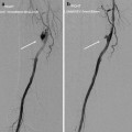
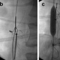
A 67-year-old man with an asymptomatic 7.3 cm AAA was referred for EVAR, and an Endurant bifurcated device (Medtronic Santa Rosa, CA) was implanted. Completion angiography confirmed successful deployment of the stent graft and the absence of any graft-related endoleak. The fabric markers were a few millimeters below the inferior margin of the renal arteries, but there appeared to be adequate apposition of fabric in the neck.
At 1-month follow-up CT scan, there was no evidence of a proximal type 1 endoleak (Fig. 16.1a
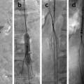
 Superficial Femoral Artery Rupture Following Angioplasty
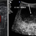 Femoral Artery Pseudoaneurysm Treated with Percutaneous Thrombin Injection
 Hemorrhage Following Percutaneous Nephrostomy
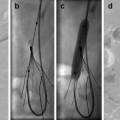
 Retrieval of a Well-Orientated IVC Filter with Embedded Struts
 Protrusion of Vena Cava Filter into the Aorta
 The Multiple Options for Retrieval of a Tilted IVC Filter


Related posts:
Superficial Femoral Artery Rupture Following Angioplasty
Femoral Artery Pseudoaneurysm Treated with Percutaneous Thrombin Injection
Hemorrhage Following Percutaneous Nephrostomy
Retrieval of a Well-Orientated IVC Filter with Embedded Struts
Protrusion of Vena Cava Filter into the Aorta
The Multiple Options for Retrieval of a Tilted IVC Filter
Stay updated, free articles. Join our Telegram channel
Full access? Get Clinical Tree
