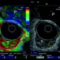
Fig. 1
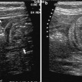
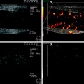
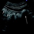
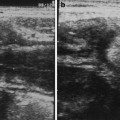
Transperineal ultrasound. Technical approach (left panel; Figures a, c) and correspondent findings (right panel, Figures b, d). With patient position is dorsal or left lateral position, the transducer is placed directly on perineal body. Transducer is pressed posteriorly and angled cranially-to-caudally, to obtain a cross sectional view, mainly in women (a, b) or placed on the perineum to obtain a long axis view (c, d). In both sections, internal anal sphincter (IAS or i) and external anal sphincter (EAS or e) appears as a hypoechoic ring and as a tissue of mixed echogenicity like in anorectal ultrasound. P: probe; R: rectum; arrow: ano-rectal angle
In this way, it is possible to obtain high-resolution images of the anal canal, the anal sphincters, pubo-rectal muscle, recto-vaginal and anovulvar septum, urinary bladder, prostate and vagina. Internal anal sphincter is the terminal condensation of the circular rectal smooth muscle. It is seen as an approximately 3-mm thick hypoechoic symmetric band (Fig. 1) sometimes demarcated from heterogeneous subepithelial tissues medially. The surrounding mixed-echogenicity striated external sphincter is slightly thicker, extends approximately 1 cm beyond the internal anal sphincter (Fig. 1


Stay updated, free articles. Join our Telegram channel

Full access? Get Clinical Tree


