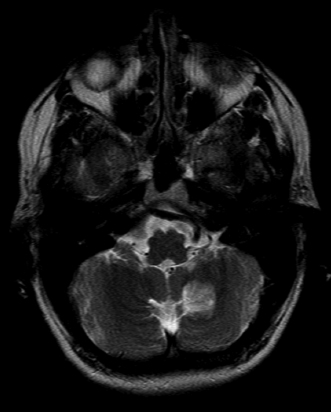
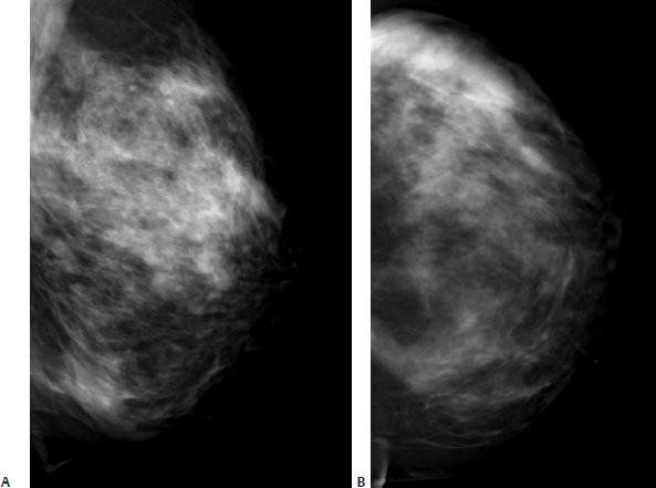
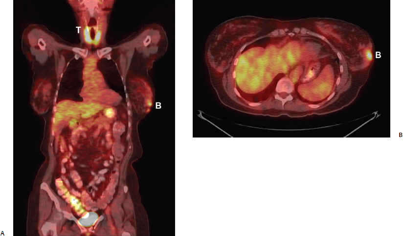
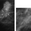
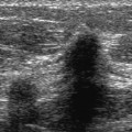
39 Unknown Primary A 60-year-old woman presents with right chin numbness. Brain MRI identifies a cerebellar mass. Because her physician suspects that the mass is a metastasis, a positron emission tomography–computed tomography (PET-CT) scan is performed, which demonstrates abnormal uptake in the left breast and thyroid. After the PET-CT scan, bilateral mammograms and left breast sonogram are performed. The sonogram identifies a cystic mass that is biopsied and found to be ductal carcinoma in situ. Bilateral breast MRI is then performed demonstrating extensive abnormal left breast enhancement. Because mastectomy is being considered, a second left breast sonogram is performed to identify lesions to biopsy that would be distant from the primary mass. The PET-CT thyroid abnormalities are also followed up with a thyroid sonogram, which identifies a suspicious right thyroid nodule. • Right chin decreased sensation • Normal breast examination • Multiple nodules in both thyroid glands Fig. 39.1 Axial T2-weighted MRI of the brain. There is a high-intensity mass in the left cerebellum. The differential diagnosis is a metastasis versus a slow-growing glioma. Fig. 39.2 After the cerebellar mass is discovered, positron emission PETCT scan is performed to identify a primary malignancy. The PET-CT demonstrates increased uptake in the thyroid (T) and the left breast (B). (A) Coronal PET-CT. (B) Axial PET-CT of chest. • Normal Fig. 39.3 Mammograms are performed as a result of the abnormal PET-CT scan. Left breast has heterogeneous breast composition without suspicious mass or calcifications. (A) Left MLO mammo-gram. (B) Left CC mammogram. Frequency (Figs. 39.4, 39.5, 39.6, and 39.7) • 14 MHz
Case 39.1: Unknown Primary
Case History
Physical Examination (Figs. 39.1 and 39.2)
Mammogram (Fig. 39.3)
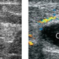
Ultrasound
Related posts:


Stay updated, free articles. Join our Telegram channel

Full access? Get Clinical Tree


