(1)
Radiology – Neuroradiology Section, S. Paolo Hospital, Bari, Italy
A 35-year-old man
3 weeks history of left-sided low back pain
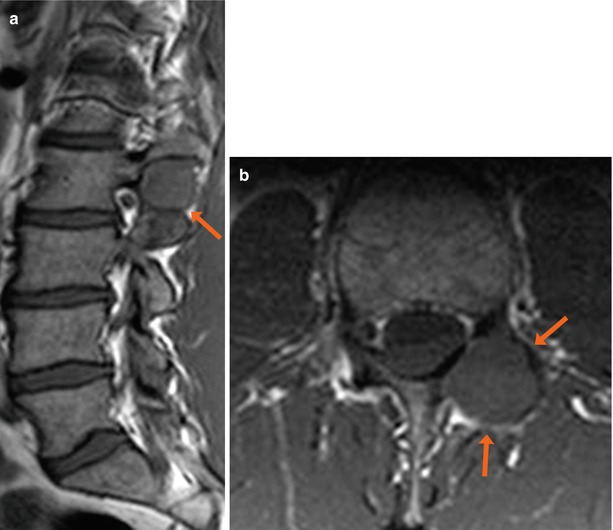
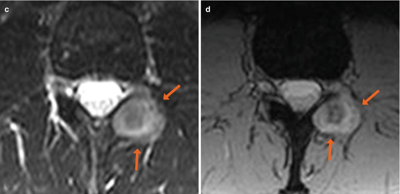
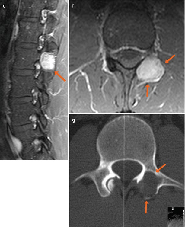
Fig. 1


Sagittal and axial SE T1-weighted image (a, b), axial TSE T2-weighted image with fat saturation (c), axial GE T2*-weighted image (d), sagittal and axial SE T1-weighted images with fat saturation following administration of contrast medium (e, f), axial CT scan (g). Extradural mass arising from the left facet joint at L2/L3 (arrow). The lesion shows intermediate-low signal intensity in T1 (a, b) and hyperintensity in T2 (c, d), with marked contrast enhancement (e, f). CT scan shows osteolysis of the same facet joint (g, arrows). The patient underwent a biopsy of the lesion, with histological diagnosis of pigmented villonodular synovitis (PVNS). PVNS is a locally aggressive proliferative disorder affecting synovium lined joints. The anatomo-pathological findings of PVNS are constituted by synovial cells lined by villous fronds containing mononuclear cells, multinucleated giant cells, fibroblasts and macrophages
Stay updated, free articles. Join our Telegram channel

Full access? Get Clinical Tree


