1 Upper Extremity
FINGERS
Points to consider
Technique
![]() Metacarpophalangeal joint must be included
Metacarpophalangeal joint must be included
![]() Always include another finger to aid identification
Always include another finger to aid identification
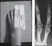
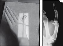
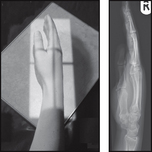
![]() AP – the fingers must be placed flat upon the cassette
AP – the fingers must be placed flat upon the cassette
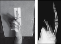
![]() Lateral – non-opaque pad can be used to help extend the finger
Lateral – non-opaque pad can be used to help extend the finger
PA (dorsipalmar) – affected finger
• Patient seated, affected side towards the X-ray table
• Palmar aspect of fingers placed on the cassette
THUMB
Points to consider
Technique
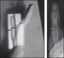
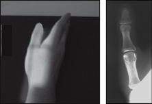
![]() AP – condyles must be equidistant from the cassette
AP – condyles must be equidistant from the cassette
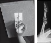
![]() Lateral – condyles must be superimposed
Lateral – condyles must be superimposed
![]() ?Trauma – consider alternative AP (trauma) projection
?Trauma – consider alternative AP (trauma) projection
AP
• Patient seated, affected side towards the X-ray table
• Thumb, elbow and shoulder at the same height (desirable but not essential)
• The hand and forearm are extended
• Hand rotated medially so that the posterior aspect of the thumb is in contact with the cassette
THUMB
HAND
Points to consider
Technique
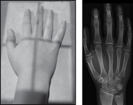
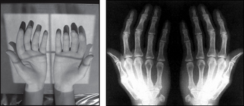
![]() Include the whole of the hand, including carpal bones and distal radius/ulna
Include the whole of the hand, including carpal bones and distal radius/ulna
![]() ?Injury confined to distal digit – limit image to that digit
?Injury confined to distal digit – limit image to that digit
![]() If you identify an injury – proceed to a lateral
If you identify an injury – proceed to a lateral
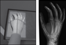
![]() PA oblique – better general assessment if fingers parallel
PA oblique – better general assessment if fingers parallel
Radiological assessment
![]() #s metacarpal neck – usually the result of a direct blow
#s metacarpal neck – usually the result of a direct blow
![]() Common site for #s – head of the fifth metacarpal – Boxer’s #
Common site for #s – head of the fifth metacarpal – Boxer’s #
![]() Look for vertical # through the base with dislocation of joint
Look for vertical # through the base with dislocation of joint
![]() Secondary ossification centres appear at age 2–3 years
Secondary ossification centres appear at age 2–3 years
![]() PA poor at showing #s of the articular surface of the metacarpal heads
PA poor at showing #s of the articular surface of the metacarpal heads
PA (dorsipalmar)

