Fig. 35.1
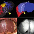
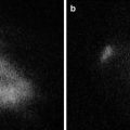
(a) Exposed abdominal cavity with xenon light source, no auto-fluorescence is detected, ureters are not visible. (b) After an average of 8 min (subjects were evaluated in the range of 5–12 min), and employing a pedal to alternate between xenon “white light” and 530 nm wavelength light sources, it was possible to visualize ureteral fluorescence in all of the animals. The distribution and location of the ureters were noted directly, with a clear distinction from the nearby vascular structures. This is especially useful for the surgeon since it provides anatomic orientation, therefore enabling visualization of real-time ureteral peristaltic flow
We were able to demonstrate that ureters can be visualized using intravenous administration of fluorescein in rats. An acceptable intensity of fluorescence was observed using a 4 mg/kg dose. Excitation of the fluorescein was noted inside the ureters only, but not in the kidney parenchyma or other retroperitoneal structures. Some of the most notable advantages of fluorescein include ease of use, low cost, and low rate of side effects at a relatively large dose. The incidence of adverse reactions is 0.6 %, which is mainly urticaria and in very few reported cases, post-injection anaphylactic shock in quick bolus [26–28].
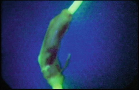
Other reports have described the use of fluorescein during open surgery [29] (Fig. 35.2).


Fig. 35.2
Ex vivo fluorescence of human ureter after nephrectomy
Conclusion
The use of a multimodal optical system that expands the surgeon’s light spectrum of view can improve surgical performance making structures clearly visible during laparoscopic and open surgery. This allows for shorter procedural duration and improved prevention and incidence of ureteral injury associated with complex pelvic surgery. Optical imaging using invisible NIR fluorescent light has several advantages over currently available intraoperative techniques. First, visualization of the ureters does not require ionizing radiation, and uses only safe wavelengths of light for ample excitation. Secondly, because fluorescence emission is invisible to the human eye, the surgical field is not stained or changed in any way. The blue dyes that are currently used stain the surgical field and have relatively poor contrast. Thirdly, imaging can be performed in real-time (up to 15 frames per second) with the merged image from the color video and NIR fluorescent cameras providing anatomical landmarks that are easily identifiable. More work is needed to identify the optimal contrast agent and light wavelength with which it is most optimally visualized. Overall, this field of knowledge is of great interest and reports a great growing potential.
References
1.
2.
3.
4.
5.
6.
Wang AC. The techniques of trocar insertion and intraoperative rethrocystoscopy in tension-free vaginal taping: an experience of 600 cases. Acta Obstet Gynecol Scand. 2004;83:293–8.CrossRefPubMed
Stay updated, free articles. Join our Telegram channel

Full access? Get Clinical Tree


