KEY FACTS
Terminology
- •
Presence of 2 separate pelvicalyceal collecting systems draining 1 kidney, which may join above bladder (partial), drain into bladder separately (complete), or beyond bladder
Imaging
- •
Asymmetric renal enlargement, 20% are bilateral
- •
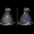
2 central echogenic renal sinuses with intervening bridging renal parenchyma
- •
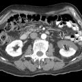
2 distinct renal pelves or 2 exiting ureters in single kidney
- •
Weigert-Meyer rule: Upper moiety ureter inserts inferior and medial to lower moiety ureter
- ○
Upper pole tends to obstruct
- ○
Lower pole tends to have vesicoureteral reflux
- ○
- •
Look for ureterocele (cystic structure) in bladder
Top Differential Diagnoses
- •
Column of Bertin
- •
Segmental multicystic dysplastic kidney
- •
Hydrosalpinx (dilated ureter in pelvis)
Pathology
- •
Abnormal bifurcation of ureteral bud
- •
Increased incidence of ureteropelvic obstruction; genital anomalies found in 50% of affected females
Clinical Issues
- •
Symptoms include infection from reflux/stasis, hematuria, abdominal/flank pain from obstruction or calculi
- •
Most often diagnosed on antenatal ultrasound or as incidental postnatal finding
- •
Longstanding obstruction, reflux and infection can lead to secondary hypertension and renal insufficiency/failure
- •
Ectopic insertion in females: Incontinence due to insertion below bladder sphincter
- •
Ectopic insertion in males: Prostatitis, epididymitis
Diagnostic Checklist
- •
Young females with recurrent urinary tract infections
- •
US, IVP or CT/MR urography are imaging modalities of choice
- •
VCUG to exclude reflux
Scanning Tips
- •
Color Doppler can be useful to distinguish collecting system from vessel in renal pelvis
- •
Ureteral jets can be helpful to identify vesicoureteral junction of both upper and lower moieties
 with hydroureter draining into a ureterocele
  . Note the upper moiety ureter inserts inferior and medial to lower moiety ureter
  .
.
 , separated by intervening bridging renal tissue.
 , reflecting a dilated upper moiety of a duplex kidney. The lower pole
  was normal.
 with dilatation of the lower moiety
  , secondary to reflux.
 compatible with duplication. The upper moiety is hydronephrotic
  and the lower moiety is also mildly dilated
  , secondary to vesicoureteral reflux.
Stay updated, free articles. Join our Telegram channel

Full access? Get Clinical Tree


