Ureteral Injury
Shaun R. Rybak
CLINICAL HISTORY
24-year-old female restrained passenger in a high-speed motor vehicle collision.
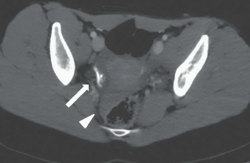 FIGURE 67A |
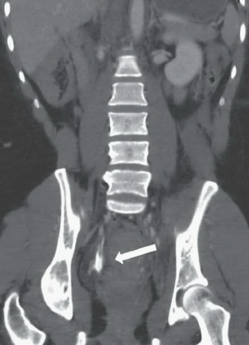 FIGURE 67B |
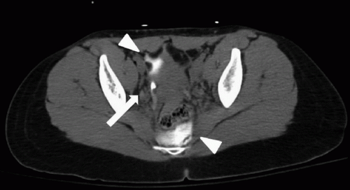 FIGURE 67C |
FINDINGS
Figures 67A and 67B: Axial and coronal contrast-enhanced CT images of the pelvis demonstrate a collection of high-density material surrounding the right distal ureter just above the level of the acetabula (arrows). The density is greater than that of the arteries. There is low- and high-density fluid in the pelvis (arrowhead), likely consisting of urine and hemorrhage. Figure 67C: Axial delayed CT image of the pelvis demonstrates increased amount of extravasated contrasted urine from the right distal ureter (arrow) in the anterior and posterior pelvis (arrowhead).
DIFFERENTIAL DIAGNOSIS
Related posts:


Stay updated, free articles. Join our Telegram channel

Full access? Get Clinical Tree


