Chapter 6. Urinary System
Patient Preparation
• No preparation is required, although if included in a complete abdomen examination, the patient may be fasting.
Equipment and Technical Factors
• A curved linear multihertz transducer is commonly used; a sector/vector transducer may be needed for intercostal imaging.
• Color Doppler imaging may be used to distinguish between vascular and nonvascular structures.
Imaging Protocol
• Longitudinal axis images through the medial, mid, and lateral aspects of each kidney are obtained; if scanning the coronal plane for the left kidney, then the anterior, mid, and inferior aspects must be documented. Echogenicity should be compared with that of the liver and spleen.
• Transverse axis images should be obtained through the superior, mid, and inferior aspects of each kidney; the transverse lateral scan plane may be used to obtain these images of the left kidney.
• A variety transducer placements (scan planes) may be used to obtain diagnostic images of the kidneys; therefore, each image must be labeled accurately to avoid confusion. A coronal scan plane provides a longitudinal and width image of the kidney.
• A variety of patient positions may be used: supine, decubitus, oblique, prone, or upright.
• Longitudinal and transverse axis images of the urinary bladder should be included in the examination; color Doppler imaging may be used to document the urine jets. The female urethra may be evaluated with transperineal imaging.
Variants
• Dromedary hump, hypertrophic column of Bertin, and extrarenal pelvis may be seen; the size of kidneys may vary with age, body habitus, sex, and state of hydration.
Sonographic Measurements
Kidneys
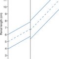
• Length: 9.0−13.0 cm
• Width (at hilum): 5.0 cm
• Depth (AP): 5.0−7.0 cm
• Volume: (L × W × D) × 0.49
• Renal sinus thickness may be measured: one half the thickness (depth, AP) of the kidney.
• Cortical thickness may be measured from the base of the pyramid to the renal capsule (approximately 1.0 cm is normal) or by subtracting the renal sinus thickness from the total kidney thickness.
Urinary bladder
• Prevoid and postvoid volume measurements may be done and a residual volume calculated; distended bladder wall measures 3.0−6.0 cm.
| Urinary System | |||
|---|---|---|---|
| Sonographic Finding(s) | Clinical Presentation | Differential Diagnosis | Next Step |
Normal to small kidney(s) with very large renal sinus Cortical thinning; cortical thickness <1.0 cm | Asymptomatic | Normal aging process Sinus lipomatosis | |
Single/multiple cysts in renal cortex Size is variable; may be very large Closely spaced cysts may appear as one large cyst with septations (“kissing cysts”) | Asymptomatic History of long-term dialysis | Development of cysts related to aging ACKD Resolved hematoma Multicystic dysplastic kidney (child) Tuberous sclerosis | “Cyst” that connects with collecting system may indicate hydronephrosis May require a renal Doppler study to demonstrate amount of viable parenchyma remaining |
Complex cystic structure arising from renal cortex (septated, internal echoes, possible calcifications) No flow with color Doppler imaging | Asymptomatic or Possible flank pain Hematuria Fever Chills Labs: WBC in urine; hematuria | Atypical cortical cyst Hemorrhagic cyst Infected cyst Abscess Tumor Prominent vessel Aneurysm Hematoma | Pus or blood clot may be present in the urinary bladder |
| Normal kidney with small, hyperechoic structure in cortex | Asymptomatic or Painless or painful hematuria (hemorrhage) Hypertension Labs: blood in urine | Angiomyolipoma (more common in females aged 40–60 years) Small RCC (incidental finding) | Power Doppler may be useful to distinguish from RCC Blood clot may be present in urinary bladder |
Hypoechoic to echogenic mass in renal cortex Variable size Large mass distorts renal shape | Asymptomatic or Possible: Hematuria Hypertension Weight loss Palpable mass Anemia Dysuria Patient on immunosuppressants or diabetic Labs: elevated BUN, creatinine, WBC | RCC Adenoma Angiomyolipoma (hamartoma) Abscess Fungal infection | Liver, contralateral kidney, IVC, renal veins, and lymph nodes may demonstrate metastases RCC tumor extension or thrombus may be found in IVC Abnormal LFT if liver metastases are present |
Normal kidney with cystic structure noted at renal hilum Connects with calyces in transverse view | Asymptomatic | Extrarenal pelvis Aneurysm Dilated proximal ureter | Lack of connection with renal pelvis contradicts diagnosis of hydronephrosis Finding collapses when patient is prone Can mimic parapelvic cyst, hydronephrosis, or calyceal diverticula |
Cystic structure noted adjacent to renal pelvis May displace renal pelvis and calyces but does not communicate with renal pelvis May mimic hydronephrosis or demonstrate concurrent hydronephrosis | Asymptomatic or Possible fever Labs: elevated BUN, creatinine (if obstructed), bacteriuria, leukocytosis | Parapelvic cyst Extrarenal pelvis Lymph node Abscess Hematoma Anechoic lymphoma | If patient is symptomatic or lymphoma is suspected, evaluate lymph nodes and urinary bladder for presence of disease Extrarenal pelvis will collapse when patient is prone |
Large kidneys with multiple irregular cysts Loss of reniform shape Remainder of kidney possibly echogenic Decreased/no visualization of renal sinus | Flank pain Dysuria Hematuria Oliguria Hypertension Palpable flank masses Labs: elevated BUN, creatinine | ADPKD ACKD | Associated with liver and pancreatic cystic disease |
Normal kidney; echogenic focus with shadowing noted in renal sinus May be multiple | Hematuria Flank pain Fever Chills Nausea Vomiting Dysuria Renal colic Hematuria Hereditary More common in males Labs: bacteriuria, blood in urine | Renal calculus (nephrolithiasis) | Clot or calculus may be present in the urinary bladder
Stay updated, free articles. Join our Telegram channel
Full access? Get Clinical Tree


|

