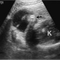
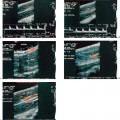
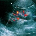
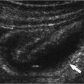
7 Urinary Tract Infections Urinary tract infection (UTI) is a common problem in childhood as well as an important cause of morbidity. Chronic or recurrent UTI can ultimately result in hypertension and renal failure, particularly when associated with obstructive uropathy or vesicoureteral reflux (VUR). Most UTIs are bacterial in origin. Diagnosis is made by documenting significant bacteriuria, defined as greater than 100,000 colony-forming units (cfu)/mL of a single organism, in one or more clean-voided specimens of urine. When the urine is obtained by suprapubic puncture or bladder catheterization, less than 100,000 cfu/mL can be considered significant.1–3 The incidence of neonatal bacteriuria has been reported as ranging from 1 to 1.4%,4–6 with a male-to-female ratio of between 2.8 and 5.4:1.7,8 The sex ratio for bacteriuria in preschool and school-age children is reversed from that seen in infancy, ranging from 0.7 to 1.9% of girls, and 0.2 to 0.4% of boys.9–13 UTI in children can be asymptomatic or symptomatic. The true incidence of UTI is unknown because of the nonspecificity or absence of symptoms in young children. The term asymptomatic bacteriuria denotes the presence of two consecutive clean-voided urine specimens, both yielding positive cultures of the same uropathogen, in a patient without urinary symptoms.14 Poor feeding, failure to thrive, irritability, vomiting, or diarrhea may be the only signs indicative of an underlying problem. Symptomatic infections may be confined to the urethra (urethritis) or bladder (cystitis), or may involve the ureter (ureteritis), the collecting system (pyelitis), or the renal parenchyma (pyelonephritis). Fever occurs in most infants with symptomatic UTIs, but is often absent in neonates.15 Once children develop speech and are toilet trained, symptoms of UTI are more readily detected. Disturbances of micturition often signal lower-tract UTI.16 Symptoms and signs of lower UTI include dysuria, frequency, and cloudy, foul-smelling urine. Pyelonephritis often results in an acute, febrile illness, associated with malaise, chills, vomiting, and flank pain. An infant with urosepsis may be critically ill, whereas in young children with less severe symptoms, it may be difficult to distinguish cystitis from pyelonephritis solely on the basis of clinical presentation. The incidence of symptomatic UTI in boys and girls under 2 years of age was shown to be the same (1.6%) in a Swedish prospective, multicenter study.17 In a study of children living in Göteborg, Sweden, published in 1974, Winberg et al estimated that the aggregate risk for symptomatic UTI up to the age of 11 years was at least 3% for girls and 1.1% for boys.15 In a more recent report from the same city, the prevalence of culture-documented, symptomatic UTIs in children up to the age of 7 years was higher than the previous estimate, with 8.4% of girls and 1.7% of boys affected.18 The majority of UTIs are caused by the family of gram-negative, aerobic bacilli known as Enterobacteriaceae that includes Escherichia, Klebsiella, Enterobacter, Citrobacter, Proteus, Providencia, Morganella, Serratia, and Salmonella species. Of these, Escherichia (E.) coli is by far the most frequently isolated organism, being responsible for ~80% of UTIs. Pseudomonas is a gram-negative, aerobic bacillus, unrelated to the Enterobacteriaceae, which may be isolated from the urine of patients with impaired immunologic defense mechanisms. The most common gram-positive organisms found in UTIs are Staphylococcus and Enterococcus. Gram-negative organisms usually infect the urinary tract by ascent from the perineum, whereas gram-positive organisms often reach the kidney hematogenously. Ascending infection occurs in ~97% of cases.19 UTI occurs when bacterial virulence factors overwhelm host resistance.20–22 Some of the most important bacterial virulence factors include adherence to uroepithelial cells, large amounts of K-antigen in the bacterial capsule, production of hemolysin and colicin, the ability to incorporate iron, and resistance to serum bactericidal activity. Bacterial adherence is an essential step in the initiation of UTI. Uropathogenic bacteria attach both to specific receptor sites on the uroepithelium and in a nonspecific fashion by electrostatic hydrophobic means. As a consequence of this attachment, virulent bacteria may ascend into the upper urinary tract in the absence of VUR or structural abnormalities. Adhesion is mediated by surface glycoprotein projections known as pili or fimbriae, which serve as ligands for glycoprotein and glycolipid receptors on uroepithelial cells. Fimbriated E. coli are categorized as either mannose-sensitive or mannose-resistant, according to their ability to agglutinate erythrocytes in the presence of mannose. E. coli expressing type I (or common) fimbriae are mannose-sensitive and are thought to be ubiquitous among UTI-causing E. coli strains. Mannose-resistant pili on E. coli can be further categorized as P- or X-fimbriated, based on their ability to agglutinate human erythrocytes expressing P (or other) blood group antigens. P-fimbriated E. coli are associated with the development of upper tract infection, probably because the major glycolipid component of renal cell membranes is the receptor for P fimbriae.23 Other bacterial virulence factors include K or capsular antigens that inhibit phagocytosis; hemolysin, which lyses phagocytes and erythrocytes resulting in the release of iron; colicin, a substance produced by some strains of E. coli that kills competing strains; and the aerobactin system, which chelates urinary iron and offers a growth advantage to bacteria that possess it.23,24 Many host resistance factors protect the urinary tract from infection, including the presence of antibodies, male sex, a low density of bacterial receptor sites on host epithelial cells, lack of perineal colonization by virulent fecal bacteria, and unimpeded flow of urine. Immunity to UTI is boosted in the neonatal period by the maternal transmission of immunoglobulin (Ig) A antibodies and other protective factors such as lactoferrin and oligosaccharides through breastfeeding that impede microbial mucosal attachment.25 The short urethra in girls appears to be the most obvious explanation for the increased relative incidence of UTIs in girls beyond the first year of life compared with boys. There is significantly increased risk of UTIs in uncircumcised male infants in the first year of life, which is believed to be due to colonization of the prepuce by uropathogenic bacteria.26–28 A genetic predisposition to childhood UTI is suggested by studies that demonstrate increased adherence of uropathogens to the uroepithelial cells of children who have had a UTI,29 as well as by a recent study that demonstrated a mutation in the neutrophil interleukin (IL)-8 receptor (CXCR1) in children with recurrent pyelonephritis.30 Frequent and complete bladder emptying eliminates bacteria and prevents UTI. The predisposition to recurrent UTIs and VUR in children with dysfunctional voiding is related to overdistention from infrequent voiding, residual volume due to inadequate emptying, and increased intravesical pressure caused by uninhibited bladder contractions. Development of normal voiding patterns in these children reduces the incidence of recurrent UTIs.31,32 A correlation has also been shown between constipation and recurrent UTIs in children.33,34 Improvement in bowel habits often results in a decrease in the incidence of recurrent UTIs. Resistance to bacterial infection may be compromised by impedance to the unidirectional flow of urine out of the urinary tract, as may occur with VUR, obstruction, or both. Obstruction can occur at any site within the urinary tract. Children with obstruction of the distal end of the ureter [either primary ureterovesical junction (UVJ) obstruction or ectopic ureterocele] or the urethra (posterior urethral valves) often present with infection. Although the most common site of obstruction of the pediatric urinary tract occurs at the ureteropelvic junction (UPJ), children with this condition do not usually present with UTI. VUR is an abnormality that is frequently associated with UTI. With VUR, urine flows in a retrograde fashion into the ureter. VUR is almost always a primary phenomenon due to incompetence of the UVJ and is not secondary to infection or obstruction.35,36 VUR is especially common in neonates and infants. It occurs in families,37 is less common in black children,38,39 and decreases in severity or resolves as the child gets older. Resolution is less likely to occur if the degree of VUR is severe or is associated with other abnormalities such as voiding dysfunction. VUR provides a pathway for bacteria within the bladder to reach the kidney. Due to the phenomenon of “aberrant micturition,” part of the urine expelled from the bladder refluxes up the ureter and flows back into the bladder. The resulting stagnation of urine encourages bacterial overgrowth.40 Secondary VUR is usually due either to an abnormality of micturition, such as dysfunctional voiding (a neurogenic bladder abnormality), or to the presence of a diverticulum at the UVJ.32,41 When VUR is present, it is the most significant host factor in the etiology of childhood pyelonephritis. The risk for acute pyelonephritis and subsequent renal scarring is related to the severity of VUR.42,43 Pyelonephritis may result in the destruction of nephrons and their replacement by fibrous tissue. Once lost, these renal units are never regenerated or replaced. The destruction of renal parenchyma is directly proportional to the severity and frequency of episodes of infection. The increased burden placed on the remaining nephrons is thought to result in hyperfiltration injury and glomerulosclerosis.44–46 The term reflux nephropathy is used to describe both the acute damage to the kidney as well as the long-term sequelae caused by reflux of infected urine.47 Chronic pyelonephritis refers to the process of fibrosis and scar formation that follows bacterial infection.48 The medulla is initially affected, but eventually the full thickness of renal parenchyma is involved. Scars are focal and frequently polar in location. When VUR and ipsilateral obstruction coexist, infection is much more common and its clinical presentation and renal sequelae tend to be more severe. This is presumably due to the fact that pressure in the collecting system is elevated and drainage of infected urine is impeded.49 Infants and young children are at higher risk than older children for the development of acute renal injury with UTI. In addition to acute morbidity, potential long-term consequences of UTI include renal scarring as well as the rarer complications of hypertension and renal failure.50 The risk for renal damage increases with recurrent UTIs.51 The significance of asymptomatic or covert bacteriuria is poorly understood. Although some authorities believe that asymptomatic children should be treated, others have postulated that the bacteria may, in fact, be nonpathogenic. These bacteria may actually protect the patient from potentially more virulent organisms by blocking epithelial receptor sites.51,52 Evaluation of children with UTI begins with a history of voiding and bowel habits in those who are toilet trained. Family history should also be obtained because heredity appears to be a factor in an individual’s predisposition to bacteriuria and VUR.37,53 Physical examination includes abdominal palpation to detect bladder distention, bowel distention from fecal impaction, and flank masses. Circumcision status is important in boys because of the increased risk of UTIs in uncircumcised boys. Abnormal findings in girls include the presence of labial adhesions or vulvovaginitis, both of which may predispose to perineal colonization by bacteria. In children with a significant history of voiding dysfunction associated with encopresis or constipation, an evaluation of perineal sensation and lower extremity reflexes and an examination of the lower back for sacral dimpling or cutaneous abnormalities suggestive of underlying spinal abnormalities should be performed. Rectal examination for fecal impaction is indicated if there is a history of encopresis or severe constipation. UTI can be diagnosed reliably only by urine culture. Thus, specimens for urine culture should be obtained in all patients in whom suspicion for infection is high. In children who are not toilet trained, an initial urine specimen is often obtained by placing a collection bag on the perineum. Although this technique has been correctly criticized for producing a high rate of contaminated specimens, the method is reliable when the culture is negative. Contamination is directly related to the length of time the bag remains on the perineum. Confirmation of all positive urine specimens obtained in this fashion is advisable prior to treatment. Bladder catheterization or suprapubic aspiration should be performed whenever the clinical situation dictates immediate treatment or confirmation of a positive urine bag specimen. In older children who are toilet trained, significant bacteriuria in a single properly collected midstream, clean-catch urine specimen is reliable for diagnostic purposes.54 Current guidelines of the American Academy of Pediatrics recommend obtaining a voiding cystourethrogram (VCUG) and a renal sonogram for infants and young children after a first febrile UTI.55 In many centers, including our own, an imaging evaluation is performed in all children who present after their first documented UTI. This approach assumes that renal scarring is both acquired and preventable, that a combination of infection and VUR is the cause of renal damage, and that high-risk cases can be clearly identified at an early stage with imaging. These assumptions have been recently challenged by reports of children with VUR and congenitally small or scarred kidneys that cannot be treated with ureteral reimplantation or prophylactic antibiotics,56,57 and at least one study that showed that treatment of VUR in childhood did not prevent end-stage renal disease attributable to reflux nephropathy.58 Another publication concluded that ultrasonography performed at the time of acute illness is of limited value, and that a VCUG for the identification of VUR is useful only if antimicrobial prophylaxis is effective in reducing reinfections and renal scarring.59 As stated by Dick and Feldman in 1996, methodologically sound, prospective studies are ultimately necessary to assess whether children with their first urinary tract infection who have routine diagnostic imaging are better off than children who have imaging for specific indications.60 Recommendations for particular radiological studies in children with culture-documented UTI are, to a certain extent, based on the availability of various imaging modalities and the experience and expertise of the imager. Most children are not studied during the acute phase of a UTI, although children hospitalized for febrile infections may be screened with ultrasound before discharge. Ultrasound is the initial examination of choice for imaging all children with a history of UTI. The role of ultrasound is to rule out urinary tract obstruction. VCUG by bladder catheterization is also performed to detect the presence of VUR. VCUG is usually reserved for children with a history of febrile UTI. Some imagers prefer a conventional, fluoroscopically monitored VCUG, whereas others prefer nuclear cystography. Conventional contrast VCUG allows more precise determination of the severity of VUR and assessment of bladder and urethral anatomy. Nuclear cystography is associated with reduced gonadal radiation and permits continuous monitoring throughout the study, which increases its sensitivity to transient VUR.61 Nuclear cystography is generally accepted to be the best examination for following children with known reflux, for documenting the result of antireflux surgery, and for screening children whose siblings have reflux.62,63 No further imaging is necessary in most cases if the cystogram and ultrasound of the kidneys and bladder are normal. Additional imaging evaluation of the upper urinary tract in children with UTI varies from institution to institution. Planar scintigraphy or single photon emission computed tomography (SPECT) of the renal parenchyma with either technetium-(Tc) 99m-labeled dimercaptosuccinic acid (DMSA) or glucoheptonate (GH) have been used to detect acute pyelonephritis and renal scarring.64–66
in Children
Epidemiology
Pathogenesis
Bacterial Virulence
Host Factors
Anatomical/Functional Abnormalities
Vesicoureteral Reflux
Clinical Outcome
Diagnostic Evaluation
Nonimaging Tests
Imaging Tests
![]()
Stay updated, free articles. Join our Telegram channel

Full access? Get Clinical Tree


