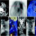
Fig. 78.1
The PET shows progression of disease due to the presence of a lung metastasis and lymph nodes characterized by increased deposition of FDG

Fig. 78.2
The CT shows a solid mass, uneven, with irregular spiculated profiles in the anterior segment of the upper lobe of the left lung invading the parietal pleura

Fig. 78.3
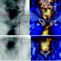 Laryngeal Squamous Carcinoma: Staging
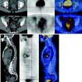 Radio-Treated Cancer of the Posterior Hemi-Circumference of the Anal Canal: Post-Actinic Fibrosis
 Bone-Destroying Metastases in Thyroid Undifferentiated Carcinoma
 Surgically Treated Endometrial Cancer: Bilateral Nodal Recurrence
 Undifferentiated Gastric Adenocarcinoma with Peritoneal Carcinosis
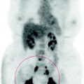
 Follow-Up of Papillary Breast Cancer: Solitary Sacroiliac Benign Lesion


PET shows high glucose consumption lesion. Presence of pleural effusion on the right contralateral to the mass
Related posts:
Laryngeal Squamous Carcinoma: Staging
Radio-Treated Cancer of the Posterior Hemi-Circumference of the Anal Canal: Post-Actinic Fibrosis
Bone-Destroying Metastases in Thyroid Undifferentiated Carcinoma
Surgically Treated Endometrial Cancer: Bilateral Nodal Recurrence
Undifferentiated Gastric Adenocarcinoma with Peritoneal Carcinosis
Follow-Up of Papillary Breast Cancer: Solitary Sacroiliac Benign Lesion
Stay updated, free articles. Join our Telegram channel
Full access? Get Clinical Tree
