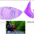
Fig. 5.1
A typical representation of a scanned cytology sample: some areas are in focus, some are not
In the second study, 22 de-identified previously screened and diagnosed cases were included, including both non-gynecological and gynecological slides [8]. Glass slides were digitalized using Aperio ScanScope XT (×20 and ×40). Three cytopathologists with and three without (3) digital experience, four cytotechnologists, and two senior pathology residents initially diagnosed the digital slides independently. Glass slides were read and recorded separately 1–3 days later. Once again, the accuracy of diagnosis and time to diagnosis were recorded. In both studies, glass slides out-performed WSI, and WSI took longer. For example, in the latter study, the diagnosis was accurate in 93 versus 86 % of cases, using glass versus digital slides. Though both techniques did well at recognizing positive cases (97.5 vs. 95.8 % for glass and digital slides, respectively), they did less well with negative cases (87.5 vs. 76.0 %, respectively), which is where the two approaches clearly differed. The average time required to read each slide also was roughly 1.5 min longer with WSI. However, the authors concluded from both studies that the problems with WSI were fixable, largely dependent upon increasing the speed of WSI capture and viewing, and generating better quality slides. They noted that only 10 % of the WSI were actually of poor quality [8] and did not comment on how many glass slides were not selected or had been tossed previously because they had been inadequate.
From these early results, what is clear is that, yes, considerable work still needs to be done to bring digital pathology to a level where it can be used with cytological specimens. On the other hand, the difference between where digital pathology is now and where it needs to be is not that great, so long as developers and investigators follow the same general validation guidelines recommended by the CAP for WSI use in standard pathology practice [8, 9].
5.2 Digital Pathology as a Teaching Tool
Chapter 3 discussed both the ease and effectiveness of using digital pathology as a teaching tool, as well as the numerous advantages. One professional school that has adopted it wholeheartedly is a school of podiatric medicine in New York.
5.2.1 New York College of Podiatric Medicine
Digital pathology, in the context of a 16-week course in basic mechanisms and systemic pathology, has now become one of the primary teaching tools at the New York College of Podiatric Medicine (NYCPM) [10]. The essential components of the digital pathology experience are (1) a laboratory in which core exercises are assigned for students to complete; (2) classroom access to computers for all students in small groups; (3) specialized software to access slides by virtual microscopy; (4) additional software that allows them to capture/download and edit slides for later presentation and sharing of observations; and (5) a course-specific (wiki) Web site (http://pathlab2014.wikifoundry.com) that allows them to enter and share their observations with every other student in the class outside of class time. For these purposes, the department’s glass slide pathology collection was converted to the VM system (Olympus) for virtual viewing.
Laboratory sessions begin with a brief introduction to the essential features of the assigned digital slides, including slides demonstrating normal histology of each tissue. The students then work in groups of four, looking at the slides at their computer stations via virtual microscopy, as well as reviewing assigned references in Wheater’s Basic Pathology and Curran’s Atlas of Histopathology, with additional major atlases also available in the room (e.g., Sandritter and Damjanov). Toward the end of each session, new slides that demonstrate the same pathology but are not among the collection they already have viewed are projected onto the students’ computer screens for open discussion across the entire class. For each slide, students are asked to volunteer their opinions about what they see, and what the likely pathology is and represents, from a disease perspective. Meanwhile, all other students are encouraged to agree or disagree with the initially expressed views and to explain why. The instructor guides the discussion with probing questions, as necessary—like “do you agree that this is subcutaneous tissue?” “do you agree that these are lymphocytes?” “do you agree that this is malignant change?” and always “why (do you agree or disagree)?” The discussion continues for each slide until a diagnostically accurate consensus is reached, aided by the instructor.
Advantages of this system are both numerous and undeniable. First, rather than four students taking brief turns examining glass slides, and perhaps not even visualizing the same field, using slides that differ from group to group, they are all able to visualize, on their computers, the same slide at the same time. Second, they can bring up images showing normal tissue and the assigned patient’s pathology, side by side, to compare and contrast them, thereby more easily identifying the areas and characteristics of normal tissue and diseased tissue. Third, the students have access not only to slides in the NYCPM collection, but from collections housed at other universities, including collections at the Mt. Sinai School of Medicine, Albert Einstein College of Medicine, Ben Gurion University, and, via the course Web site (wiki), the University of Indiana and University of Iowa. Fourth, using programs like Paint, Image Manager, and Adobe, they have the capability to modify each slide—for example, to cut and paste significant areas, and to annotate, underline, circle, or add arrows; however, they wish to preserve their value-added versions of each slide. Fifth, via the online wiki Web site to which all students have 24/7 access, they can enter their personally modified slides and make them available to all, along with their comments and annotations. As such, all slides can be shared by all; modified and improved by all; downloaded and saved by all, either singly or as part of their own illustrated MS-Word notes or PowerPoint presentations; and, ultimately, studied by all.
5.2.2 Universal Education
Yet another advantage of a digital reference collection is that this digital educational tool can be used off-site, no matter the distance from the home university where it was created. All that is needed for those wishing to use it is to have computer access and have been allowed to register for the course Web site.
In the survey of the literature we conducted while writing this book, we found that education plays a role in virtually all digital pathology setups. Whether it is for sharing information with colleagues, or the formal offering of a course (as in the case of NYCPM), or in the training of pathology residents, or in continuing medical education for pathologists already in practice, it makes sense to use the infrastructure in place and get as much use out of it as possible.
Continuing medical education is an extremely important application, for a variety of reasons. For example, rare tumors are a fact of life, and in rural areas or small countries they may only appear once every so many years. They also might not be observed at all during a pathologist’s formal years of training. It is useful, therefore, for practicing pathologists to be able to regularly update their skills by viewing pathology on their computer screen. The pathology can come from a patient who lives halfway around the world but who has a rare lesion of interest. Education then goes together with the construction of biobanks (see Chap. 3), whereby one can use the biobank as a study tool to stay current in diagnostic protocols.
This brings us back to telepathology, for which there also is an undeniable educational component. Initially, set up to assist in areas where pathology services are sparsely available, it stands to reason that the institute that is on the submitting end of the telepathology workflow uses the information it receives back, in terms of primary diagnoses and second opinions, to update its own knowledge base/skill set and pass it on to its associates and affiliates.
5.3 Telepathology in Developing Countries
One of the harsh social realities of life on Earth is that huge discrepancies exist in the standard of living and access to technical advances in different countries and regions. And in no field is this observed more than in the field of Medicine. For example, whereas almost 8,000 magnetic resonance image (MRI) scanners serve a US population of less than 320 million, in India, there are only about 600 units serving more than 1.2 billion people [11–13


Stay updated, free articles. Join our Telegram channel

Full access? Get Clinical Tree


