and Clarisse Dromain2
(1)
Department of Radiology, San Giovanni Hospital, Roma, Italy
(2)
Department of Radiology, Institut de Cancerologie Gustav Roussy, VilleJuif – Paris, France
Abstract
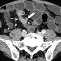
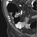
Vasculitis can cause local or diffuse pathological changes in the gastrointestinal tract, resulting in nonspecific paralytic ileus, mesenteric ischemia, submucosal edema and hemorrhage, or bowel perforation or stricture.
Vasculitis, GI Tract
Vasculitis can cause local or diffuse pathological changes in the gastrointestinal tract, resulting in nonspecific paralytic ileus, mesenteric ischemia, submucosal edema and hemorrhage, or bowel perforation or stricture.
The extent and clinical course of disease depend on the size and location of the affected vessel and the histological characteristics of the lesion. Vasculitis may primarily involve large vessels (e.g., giant cell arteritis, Takayasu arteritis), medium-sized vessels (e.g., polyarteritis nodosa, Kawasaki disease, primary granulomatous central nervous system vasculitis), or small vessels (e.g., Wegener granulomatosis, Churg–Strauss syndrome, microscopic polyangiitis, Henoch–Schönlein syndrome, systemic lupus erythematosus, rheumatoid vasculitis, Behçet syndrome). Most of them may show gastrointestinal involvement.
Radiologic findings in various types of vasculitis often overlap considerably and therefore have limited value in making a specific diagnosis. Nevertheless, the possibility of vasculitis should be considered whenever mesenteric ischemic changes occur in young patients, are noted at unusual sites (e.g., stomach, duodenum, rectum), and have a tendency to concomitantly involve the small and large intestine and when differential diagnosis with more common disease presenting with similar findings is ruled out.
Therefore clinical findings and lab test as well as biopsy and histology on target organ are crucial to reach the correct diagnosis.
In polyarteritis nodosa the kidneys are the most common involved organs: however, gastrointestinal tract and more commonly the small bowel is the second target of this necrotizing vasculitis. Imaging findings overlap with those of acute mesenteric ischemia, and multiple mesenteric renal, hepatic, and splenic pseudoaneurysm can be detected using CT angiography or more easily with conventional angiography.
In Wegener granulomatosis the target organs are represented by the lung and airways, although intestinal manifestation mimics inflammatory bowel disease.
< div class='tao-gold-member'>
Only gold members can continue reading. Log In or Register to continue

Stay updated, free articles. Join our Telegram channel

Full access? Get Clinical Tree


