KEY FACTS
Terminology
- •
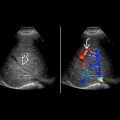
Dilatation of pampiniform plexus > 2-3 mm due to congestion and retrograde flow in internal spermatic vein
Imaging
- •
Dilated serpiginous veins at superior pole of testis
- •
“Flash” of color Doppler with Valsalva
- •
Left (78%), right (6%), bilateral (16%)
- •
Varicose veins > 2-3 mm diameter, increase in size with Valsalva
Pathology
- •
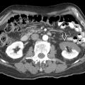
Primary: Incompetent venous valve near junction of left renal vein (LRV) and IVC
- •
Secondary: Obstruction of LRV by renal or adrenal tumor, nodes or rarely SMA compression
Clinical Issues
- •
Most frequent cause of male infertility
- •
Vague scrotal discomfort or pressure when standing
- •
10-15% of men in USA have varicoceles
- •
Subclinical varicocele in 40-75% of infertile men
- •
Catheter embolization, surgical treatment, or sclerotherapy if symptomatic
- •
Emerging research suggests even subclinical varicoceles should be treated
Diagnostic Checklist
- •
Consider left renal vein occlusion by tumor in elderly male patient presenting with recent onset of varicocele
- •
Varicocele diagnosed when vessel > 2 mm during quiet respiration in supine position
Scanning Tips
- •
Unilateral and left-sided varicoceles common; isolated right-sided varicoceles uncommon and should prompt evaluation along IVC to exclude mass
- •
Valsalva with color Doppler essential for diagnosis of small varicoceles
- •
Measure diameter of vein on grayscale; blooming artifact on color Doppler will obscure true diameter and lead to overestimation of diameter
- •
Measure vein from inner wall to inner wall
- •
Avoid mistaking vas deferens as dilated vein
- •
Varicoceles usually lie superior to testicle but may lie posterior or lateral to testicle
 in the spermatic cord and along the posterosuperior aspect of the testis
  .
.
Stay updated, free articles. Join our Telegram channel

Full access? Get Clinical Tree


