Vascular Calcifications and Aneurysms
Michael P. Federle, MD, FACR
Kathleen E. Jacobs, BA
Key Facts
Terminology
Arterial calcifications are usually indicative of atherosclerosis or diabetic vasculopathy
Venous calcifications are thrombi or result of venous (usually portal) hypertension
Imaging
Ring-like or “tram-track” linear densities in vessel walls
“Conduit” calcification (implies calcification in wall of hollow tube)
Iliac artery aneurysm
Substantial danger of rupture with diameter > 5 cm
Visceral artery aneurysms
Splenic: Most common visceral aneurysm (60%)
Danger of rupture if large, in pregnant patient, or in patient with advanced liver disease
May mimic pancreatic tumor
Renal artery aneurysm
May mimic hypervascular or hypovascular tumor
Phleboliths (“vein stones”)
More common in multiparous women
Below level of ureterovesical junction
Radiolucent center with smooth spherical outline
Portal venous calcification
Usually indicates chronic ↑ pressure (portal hypertension)
Portal vein aneurysm may mimic pancreatic or other abdominal mass
Top Differential Diagnoses
Ureteral calculus
Renal calculi
Mucinous cystic pancreatic tumor
Pancreatic islet cell tumors
Renal cell carcinoma
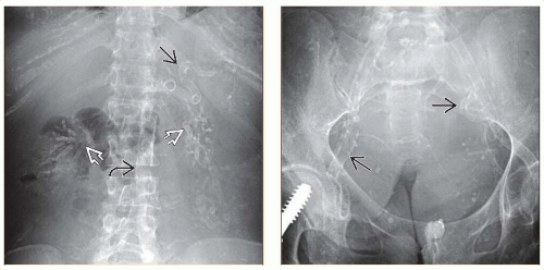 (Left) Radiograph in a 50-year-old woman with type 1 diabetes shows severe, diffuse calcification of the aortic walls  & medium-sized vessels, including the splenic & medium-sized vessels, including the splenic  & renal arteries & renal arteries  . This pattern of long, continuous calcification (Mönckeberg) is almost pathognomonic of diabetic angiopathy. (Right) Pelvic radiograph in the same patient. Mönckeberg calcification . This pattern of long, continuous calcification (Mönckeberg) is almost pathognomonic of diabetic angiopathy. (Right) Pelvic radiograph in the same patient. Mönckeberg calcification  does not cause significant luminal narrowing, unlike atherosclerotic calcified plaques. does not cause significant luminal narrowing, unlike atherosclerotic calcified plaques. |
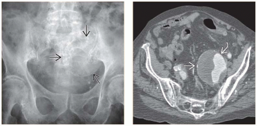 (Left) Plain radiograph shows a large, peripherally calcified pelvic mass  that raises concern for a possible neoplasm. (Right) Axial CECT in the same patient shows that the mass is a large iliac artery aneurysm. Note the enhancement that raises concern for a possible neoplasm. (Right) Axial CECT in the same patient shows that the mass is a large iliac artery aneurysm. Note the enhancement  of a portion of the lumen and the unenhanced thrombus of a portion of the lumen and the unenhanced thrombus  . . |
TERMINOLOGY
Definitions
Arterial calcifications are usually indicative of atherosclerosis or diabetic vasculopathy
Venous calcifications are thrombi or result from venous (usually portal) hypertension
Aneurysm implies significant dilation of vessel lumen with intact endothelial lining
Pseudoaneurysm = dilated lumen without intact lining (e.g., result of arterial injury from trauma, pancreatitis, etc.)
IMAGING
General Features
Best diagnostic clue
Ring-like or “tram-track” linear calcifications
“Conduit” calcification = calcification in wall of hollow tube
Location
Abdomen ± pelvis
Size
Few mm to several cm
Morphology
Arterial calcification ± aneurysm
Ring-like calcification on cross sectional view
“Tram-track” linear calcifications on long axis
Peripheral, oblong, or “eggshell” calcification with aneurysm
Exceeds diameter of normal vessel
Often discontinuous
Commonly in aorta and in abdominal and pelvic vessels
Focal discontinuity in circumferential wall calcifications is more commonly observed on CT in unstable or ruptured aneurysms (especially aortic)
Useful to observe changes in continuity of calcification with time
Calcified small arteries = string-like appearance
Uterine artery: Horizontal or slightly undulating linear opacities (often in diabetic women)Related posts:

Stay updated, free articles. Join our Telegram channel

Full access? Get Clinical Tree


